


 癌症的基本特征包括细胞增殖、血管生成、迁移、凋亡逃避机制和细胞永生等。找到癌症发生过程中这些通路的关键标记物和对应的抗体用于检测至关重要。
 为您推荐一个泛素化位点预测神器——泛素化分析工具,可以为您的蛋白的泛素化位点作出预测和评分。
 细胞自噬受体图形绘图工具为你的蛋白的细胞受体结合位点作出预测和评分,识别结合到自噬通路中的蛋白是非常重要的,便于让我们理解自噬在正常生理、病理过程中的作用,如发育、细胞分化、神经退化性疾病、压力条件下、感染和癌症。
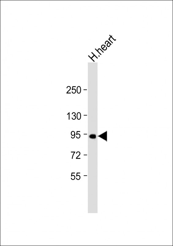
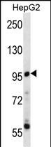
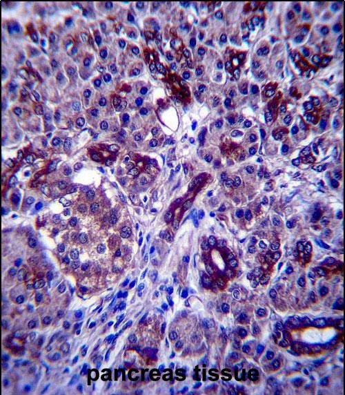
PPP1R3F Antibody (C-term)
Affinity Purified Rabbit Polyclonal Antibody (Pab)
- 产品详情
- 实验流程
- 背景知识
Application
| WB, IHC-P, E |
|---|---|
| Primary Accession | Q6ZSY5 |
| Other Accession | Q9JIG4, NP_149992.3 |
| Reactivity | Human |
| Predicted | Mouse |
| Host | Rabbit |
| Clonality | Polyclonal |
| Isotype | Rabbit IgG |
| Calculated MW | 82798 Da |
| Antigen Region | 549-578 aa |
| Gene ID | 89801 |
|---|---|
| Other Names | Protein phosphatase 1 regulatory subunit 3F, R3F, PPP1R3F |
| Target/Specificity | This PPP1R3F antibody is generated from rabbits immunized with a KLH conjugated synthetic peptide between 549-578 amino acids from the C-terminal region of human PPP1R3F. |
| Dilution | WB~~1:1000 IHC-P~~1:100~500 E~~Use at an assay dependent concentration. |
| Format | Purified polyclonal antibody supplied in PBS with 0.09% (W/V) sodium azide. This antibody is purified through a protein A column, followed by peptide affinity purification. |
| Storage | Maintain refrigerated at 2-8°C for up to 2 weeks. For long term storage store at -20°C in small aliquots to prevent freeze-thaw cycles. |
| Precautions | PPP1R3F Antibody (C-term) is for research use only and not for use in diagnostic or therapeutic procedures. |
| Name | PPP1R3F |
|---|---|
| Function | Glycogen-targeting subunit for protein phosphatase 1 (PP1). |
| Cellular Location | Membrane; Single-pass membrane protein |
| Tissue Location | Expressed in brain, skeletal muscle and heart. |
For Research Use Only. Not For Use In Diagnostic Procedures.
Provided below are standard protocols that you may find useful for product applications.
BACKGROUND
This gene encodes a protein that has been identified as one of several type-1 protein phosphatase (PP1) regulatory subunits. One or two of these subunits, together with the well-conserved catalytic subunit, can form the PP1 holoenzyme, where the regulatory subunit functions to regulate substrate specificity and/or targeting to a particular cellular compartment. Alternative splicing results in multiple transcript variants.
REFERENCES
Suttner, K., et al. J. Allergy Clin. Immunol. 125(6):1395-1399(2010)
Piton, A., et al. Mol. Psychiatry (2010) In press :
Ceulemans, H., et al. Bioessays 24(4):371-381(2002)
Schindelhauer, D., et al. Genome Res. 6(11):1056-1069(1996)
终于等到您。ABCEPTA(百远生物)抗体产品。
点击下方“我要评价 ”按钮提交您的反馈信息,您的反馈和评价是我们最宝贵的财富之一,
我们将在1-3个工作日内处理您的反馈信息。
如有疑问,联系:0512-88856768 tech-china@abcepta.com.


