


 癌症的基本特征包括细胞增殖、血管生成、迁移、凋亡逃避机制和细胞永生等。找到癌症发生过程中这些通路的关键标记物和对应的抗体用于检测至关重要。
 为您推荐一个泛素化位点预测神器——泛素化分析工具,可以为您的蛋白的泛素化位点作出预测和评分。
 细胞自噬受体图形绘图工具为你的蛋白的细胞受体结合位点作出预测和评分,识别结合到自噬通路中的蛋白是非常重要的,便于让我们理解自噬在正常生理、病理过程中的作用,如发育、细胞分化、神经退化性疾病、压力条件下、感染和癌症。
UBE2D2 Antibody (C-term)
Affinity Purified Rabbit Polyclonal Antibody (Pab)
- 产品详情
- 实验流程
- 背景知识
Application
| WB, E |
|---|---|
| Primary Accession | P62837 |
| Other Accession | P62840, P62839, P62838, Q1RMX2, NP_003330.1, NP_862821.1 |
| Reactivity | Human |
| Predicted | Bovine, Mouse, Rat, Xenopus |
| Host | Rabbit |
| Clonality | Polyclonal |
| Isotype | Rabbit IgG |
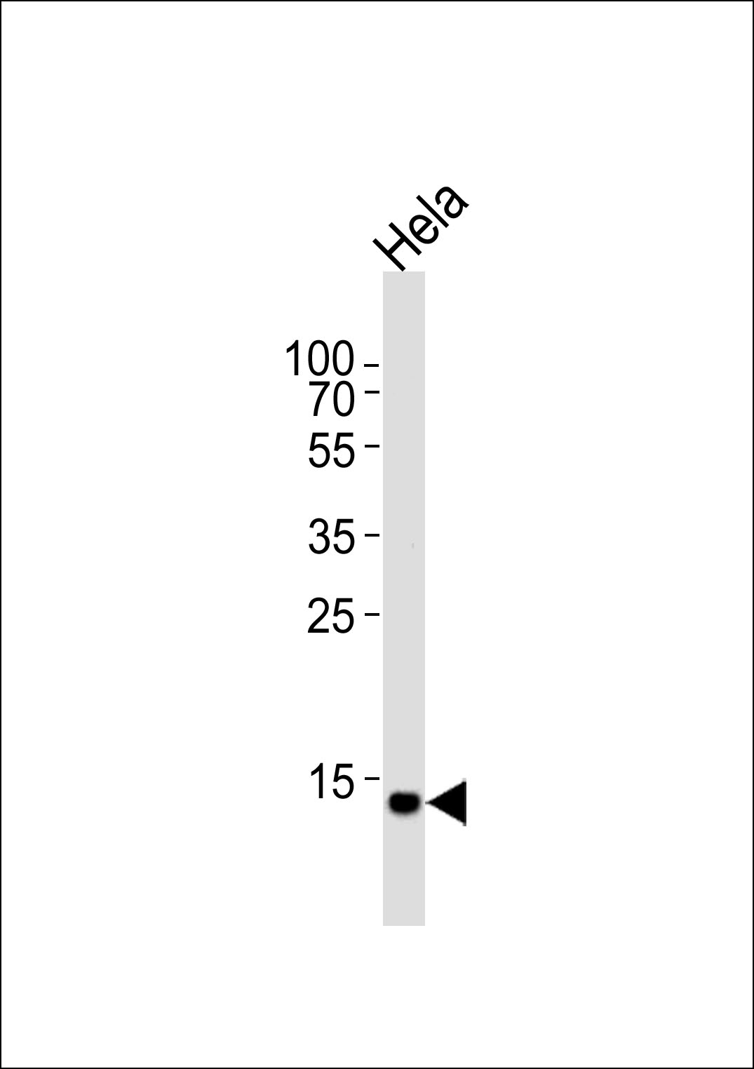
| Calculated MW | 16735 Da |
| Antigen Region | 112-140 aa |
| Gene ID | 7322 |
|---|---|
| Other Names | Ubiquitin-conjugating enzyme E2 D2, Ubiquitin carrier protein D2, Ubiquitin-conjugating enzyme E2(17)KB 2, Ubiquitin-conjugating enzyme E2-17 kDa 2, Ubiquitin-protein ligase D2, p53-regulated ubiquitin-conjugating enzyme 1, UBE2D2, PUBC1, UBC4, UBC5B, UBCH4, UBCH5B |
| Target/Specificity | This UBE2D2 antibody is generated from rabbits immunized with a KLH conjugated synthetic peptide between 112-140 amino acids from the C-terminal region of human UBE2D2. |
| Dilution | WB~~1:1000 E~~Use at an assay dependent concentration. |
| Format | Purified polyclonal antibody supplied in PBS with 0.09% (W/V) sodium azide. This antibody is purified through a protein A column, followed by peptide affinity purification. |
| Storage | Maintain refrigerated at 2-8°C for up to 2 weeks. For long term storage store at -20°C in small aliquots to prevent freeze-thaw cycles. |
| Precautions | UBE2D2 Antibody (C-term) is for research use only and not for use in diagnostic or therapeutic procedures. |
| Name | UBE2D2 |
|---|---|
| Synonyms | PUBC1, UBC4, UBC5B, UBCH4, UBCH5B |
| Function | Accepts ubiquitin from the E1 complex and catalyzes its covalent attachment to other proteins (PubMed:10329681, PubMed:18042044, PubMed:18703417, PubMed:20061386, PubMed:20403326, PubMed:20525694, PubMed:26475854, PubMed:28322253). Catalyzes 'Lys-48'- linked polyubiquitination (PubMed:10329681, PubMed:18042044, PubMed:18359941, PubMed:18703417, PubMed:20061386, PubMed:20403326, PubMed:20525694, PubMed:26475854). Mediates the selective degradation of short-lived and abnormal proteins (PubMed:10329681, PubMed:18042044, PubMed:18359941, PubMed:18703417, PubMed:20061386, PubMed:20403326, PubMed:20525694, PubMed:26475854). Functions in the E6/E6-AP-induced ubiquitination of p53/TP53 (PubMed:15280377). Mediates ubiquitination of PEX5 and SQSTM1 and autoubiquitination of STUB1 and TRAF6 (PubMed:18359941, PubMed:28322253). Involved in the signal-induced conjugation and subsequent degradation of NFKBIA, FBXW2-mediated GCM1 ubiquitination and degradation, MDM2-dependent degradation of p53/TP53 and the activation of MAVS in the mitochondria by RIGI in response to viral infection (PubMed:18703417, PubMed:20403326). Essential for viral activation of IRF3 (PubMed:19854139). |
For Research Use Only. Not For Use In Diagnostic Procedures.
Provided below are standard protocols that you may find useful for product applications.
BACKGROUND
The modification of proteins with ubiquitin is an important cellular mechanism for targeting abnormal or short-lived proteins for degradation. Ubiquitination involves at least three classes of enzymes: ubiquitin-activating enzymes, or E1s, ubiquitin-conjugating enzymes, or E2s, and ubiquitin-protein ligases, or E3s. This gene encodes a member of the E2 ubiquitin-conjugating enzyme family. This enzyme functions in the ubiquitination of the tumor-suppressor protein p53, which is induced by an E3 ubiquitin-protein ligase. Two alternatively spliced transcript variants have been found for this gene and they encode distinct isoforms.
REFERENCES
Bailey, S.D., et al. Diabetes Care 33(10):2250-2253(2010)
Wu, K., et al. Mol. Cell 37(6):784-796(2010)
Vina-Vilaseca, A., et al. J. Biol. Chem. 285(10):7645-7656(2010)
Sakata, E., et al. Structure 18(1):138-147(2010)
Kamadurai, H.B., et al. Mol. Cell 36(6):1095-1102(2009)
终于等到您。ABCEPTA(百远生物)抗体产品。
点击下方“我要评价 ”按钮提交您的反馈信息,您的反馈和评价是我们最宝贵的财富之一,
我们将在1-3个工作日内处理您的反馈信息。
如有疑问,联系:0512-88856768 tech-china@abcepta.com.


