


 癌症的基本特征包括细胞增殖、血管生成、迁移、凋亡逃避机制和细胞永生等。找到癌症发生过程中这些通路的关键标记物和对应的抗体用于检测至关重要。
 为您推荐一个泛素化位点预测神器——泛素化分析工具,可以为您的蛋白的泛素化位点作出预测和评分。
 细胞自噬受体图形绘图工具为你的蛋白的细胞受体结合位点作出预测和评分,识别结合到自噬通路中的蛋白是非常重要的,便于让我们理解自噬在正常生理、病理过程中的作用,如发育、细胞分化、神经退化性疾病、压力条件下、感染和癌症。
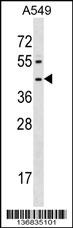
FPR1 Antibody (Center)
Affinity Purified Rabbit Polyclonal Antibody (Pab)
- 产品详情
- 实验流程
- 背景知识
Application
| WB, E |
|---|---|
| Primary Accession | P21462 |
| Other Accession | NP_002020.1, NP_001180235.1 |
| Reactivity | Human |
| Host | Rabbit |
| Clonality | Polyclonal |
| Isotype | Rabbit IgG |
| Calculated MW | 38446 Da |
| Antigen Region | 165-193 aa |
| Gene ID | 2357 |
|---|---|
| Other Names | fMet-Leu-Phe receptor, fMLP receptor, N-formyl peptide receptor, FPR, N-formylpeptide chemoattractant receptor, FPR1 |
| Target/Specificity | This FPR1 antibody is generated from rabbits immunized with a KLH conjugated synthetic peptide between 165-193 amino acids from the Central region of human FPR1. |
| Dilution | WB~~1:1000 E~~Use at an assay dependent concentration. |
| Format | Purified polyclonal antibody supplied in PBS with 0.09% (W/V) sodium azide. This antibody is purified through a protein A column, followed by peptide affinity purification. |
| Storage | Maintain refrigerated at 2-8°C for up to 2 weeks. For long term storage store at -20°C in small aliquots to prevent freeze-thaw cycles. |
| Precautions | FPR1 Antibody (Center) is for research use only and not for use in diagnostic or therapeutic procedures. |
| Name | FPR1 {ECO:0000303|PubMed:25109685} |
|---|---|
| Function | Pattern recognition G-protein coupled receptor (PRR/GPCR) involved in innate recognition of N-formyl-methionyl peptides derived from invading microbes and host mitochondria as pathogen- and damage- associated molecular patterns (PAMPs and DAMPs). Functions as a sensor of PAMPs and DAMPs released upon microbial infection or tissue damage, triggering immune cell activation and chemotaxis to eliminate pathogens and restore tissue homeostasis (PubMed:24108355, PubMed:25605714, PubMed:35217703, PubMed:36064945). Peptide binding leads to conformational changes coupled to heterotrimeric G(i) protein signaling. Upon GDP to GTP conversion, G(i)-alpha subunit dissociates from G-beta and G-gamma subunits. Free G(i)-alpha subunit inhibits cyclic adenylate cyclase and cAMP synthesis whereas the G-beta and G- gamma dimer activates downstream phospholipase C-beta and phosphoinositide 3-kinase signaling cascades leading to Ca(2+) influx (PubMed:10514456, PubMed:15153520, PubMed:1712023, PubMed:25605714, PubMed:35217703, PubMed:36064945). Displays two affinity states for peptide agonists, low and high, likely accounting for selective signaling of myeloid cell functions at different phases of the inflammatory response. Subnanomolar concentrations of peptide agonists induce myeloid cell chemotaxis, whereas micromolar concentrations trigger degranulation and superoxide production (PubMed:2161213, PubMed:2176894, PubMed:24108355, PubMed:25605714). May recognize a myriad of bacterial signal peptides indicative of an evolutionary conserved detection mechanism in host defense against bacterial infection. Triggers bactericidal functions of neutrophils and phagocytes in response to N-formyl-Met-Leu-Phe (fMLF) which is part of the signal peptide sequences of hundreds distinct bacterial strains (PubMed:25605714). In the homeostatic wound healing response to tissue injury, senses 'necrotaxis' DAMP-type signals released in the form of mitochondria-derived N-formylated peptides and guides neutrophil trafficking toward necrotic cells within the injury site (By similarity). In the context of antitumor immunity, interacts with ANXA1 and guides dendritic cell positioning in close proximity to necrotic tumor cells, allowing for tumor-associated antigen uptake and cross- presentation to T cells (PubMed:24108355, PubMed:26516201). Receptor for TAFA4, mediates its effects on chemoattracting macrophages, promoting phagocytosis and increasing reactive oxygen species (ROS) release (PubMed:25109685). Receptor for cathepsin CTSG, leading to increased phagocyte chemotaxis (PubMed:15210802). Beyond canonical N- terminal formylated peptide agonists, can also be activated by C- terminal amidated peptides, which appear to all share a tripartite structure motif oriented around a carboxyl group (PubMed:24108355, PubMed:25605714). Differential signaling is also defined by receptor oligomerization state. Pro-resolving ligands, such as lipoxin A4 or ANXA1, induce the formation of FPR1:FPR2 heterodimers triggering proapoptotic JNK pathway in neutrophils (PubMed:24108355). |
| Cellular Location | Cell membrane; Multi-pass membrane protein. Note=Internalizes in presence of its ligands, fMLP, TAFA4 and CTSG. |
| Tissue Location | Monocytes (at protein level) (PubMed:25605714). Neutrophils. |
For Research Use Only. Not For Use In Diagnostic Procedures.
Provided below are standard protocols that you may find useful for product applications.
BACKGROUND
This gene encodes a G protein-coupled receptor of mammalian phagocytic cells that is a member of the G-protein coupled receptor 1 family. The protein mediates the response of phagocytic cells to invasion of the host by microorganisms and is important in host defense and inflammation.
REFERENCES
Davila, S., et al. Genes Immun. 11(3):232-238(2010)
Huang, J., et al. Br. J. Cancer 102(6):1052-1060(2010)
Segat, L., et al. Vaccine 28(10):2201-2206(2010)
Zhu, X.L., et al. Beijing Da Xue Xue Bao 41(6):664-668(2009)
Kobayashi, T., et al. J. Dent. Res. 88(12):1137-1141(2009)
终于等到您。ABCEPTA(百远生物)抗体产品。
点击下方“我要评价 ”按钮提交您的反馈信息,您的反馈和评价是我们最宝贵的财富之一,
我们将在1-3个工作日内处理您的反馈信息。
如有疑问,联系:0512-88856768 tech-china@abcepta.com.


