


 癌症的基本特征包括细胞增殖、血管生成、迁移、凋亡逃避机制和细胞永生等。找到癌症发生过程中这些通路的关键标记物和对应的抗体用于检测至关重要。
 为您推荐一个泛素化位点预测神器——泛素化分析工具,可以为您的蛋白的泛素化位点作出预测和评分。
 细胞自噬受体图形绘图工具为你的蛋白的细胞受体结合位点作出预测和评分,识别结合到自噬通路中的蛋白是非常重要的,便于让我们理解自噬在正常生理、病理过程中的作用,如发育、细胞分化、神经退化性疾病、压力条件下、感染和癌症。
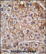
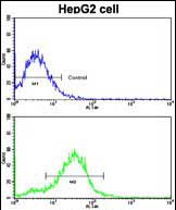
BMPR2 Antibody (N-term)
Purified Rabbit Polyclonal Antibody (Pab)
- 产品详情
- 实验流程
- 背景知识
Application
| WB, IHC-P, FC, E |
|---|---|
| Primary Accession | Q13873 |
| Other Accession | O35607 |
| Reactivity | Human, Rat, Mouse |
| Host | Rabbit |
| Clonality | Polyclonal |
| Isotype | Rabbit IgG |
| Calculated MW | 115201 Da |
| Antigen Region | 27-56 aa |
| Gene ID | 659 |
|---|---|
| Other Names | Bone morphogenetic protein receptor type-2, BMP type-2 receptor, BMPR-2, Bone morphogenetic protein receptor type II, BMP type II receptor, BMPR-II, BMPR2, PPH1 |
| Target/Specificity | This BMPR2 antibody is generated from rabbits immunized with a KLH conjugated synthetic peptide between 27~56 amino acids from the N-terminal region of human BMPR2. |
| Dilution | WB~~1:1000 IHC-P~~1:100~500 FC~~1:10~50 E~~Use at an assay dependent concentration. |
| Format | Purified polyclonal antibody supplied in PBS with 0.09% (W/V) sodium azide. This antibody is prepared by Saturated Ammonium Sulfate (SAS) precipitation followed by dialysis against PBS. |
| Storage | Maintain refrigerated at 2-8°C for up to 2 weeks. For long term storage store at -20°C in small aliquots to prevent freeze-thaw cycles. |
| Precautions | BMPR2 Antibody (N-term) is for research use only and not for use in diagnostic or therapeutic procedures. |
| Name | BMPR2 |
|---|---|
| Synonyms | PPH1 |
| Function | On ligand binding, forms a receptor complex consisting of two type II and two type I transmembrane serine/threonine kinases. Type II receptors phosphorylate and activate type I receptors which autophosphorylate, then bind and activate SMAD transcriptional regulators. Can also mediate signaling through the activation of the p38MAPK cascade (PubMed:12045205). Binds to BMP7, BMP2 and, less efficiently, BMP4. Binding is weak but enhanced by the presence of type I receptors for BMPs. Mediates induction of adipogenesis by GDF6. Promotes signaling also by binding to activin A/INHBA (PubMed:24018044). |
| Cellular Location | Cell membrane; Single-pass type I membrane protein |
| Tissue Location | Highly expressed in heart and liver. |
For Research Use Only. Not For Use In Diagnostic Procedures.
Provided below are standard protocols that you may find useful for product applications.
BACKGROUND
BMPR2 is a member of the bone morphogenetic protein (BMP) receptor family of transmembrane serine/threonine kinases. The ligands of this receptor are BMPs, which are members of the TGF-beta superfamily. BMPs are involved in endochondral bone formation and embryogenesis. These proteins transduce their signals through the formation of heteromeric complexes of 2 different types of serine (threonine) kinase receptors: type I receptors of about 50-55 kD and type II receptors of about 70-80 kD. Type II receptors bind ligands in the absence of type I receptors, but they require their respective type I receptors for signaling, whereas type I receptors require their respective type II receptors for ligand binding. Mutations in BMPR2 have been associated with primary pulmonary hypertension.
REFERENCES
Pouliot, F., et al., Cancer Res. 63(2):277-281 (2003).
Nishihara, A., et al., Mol. Biol. Cell 13(9):3055-3063 (2002).
Humbert, M., et al., Eur Respir J 20(3):518-523 (2002).
Vitt, U.A., et al., Biol. Reprod. 67(2):473-480 (2002).
Nohe, A., et al., J. Biol. Chem. 277(7):5330-5338 (2002).
终于等到您。ABCEPTA(百远生物)抗体产品。
点击下方“我要评价 ”按钮提交您的反馈信息,您的反馈和评价是我们最宝贵的财富之一,
我们将在1-3个工作日内处理您的反馈信息。
如有疑问,联系:0512-88856768 tech-china@abcepta.com.


