


 癌症的基本特征包括细胞增殖、血管生成、迁移、凋亡逃避机制和细胞永生等。找到癌症发生过程中这些通路的关键标记物和对应的抗体用于检测至关重要。
 为您推荐一个泛素化位点预测神器——泛素化分析工具,可以为您的蛋白的泛素化位点作出预测和评分。
 细胞自噬受体图形绘图工具为你的蛋白的细胞受体结合位点作出预测和评分,识别结合到自噬通路中的蛋白是非常重要的,便于让我们理解自噬在正常生理、病理过程中的作用,如发育、细胞分化、神经退化性疾病、压力条件下、感染和癌症。
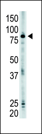
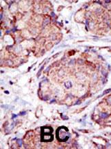
SMURF2 Antibody (N-term)
Purified Rabbit Polyclonal Antibody (Pab)
- 产品详情
- 文献引用 : 2
- 实验流程
- 背景知识
Application
| WB, IHC-P, E |
|---|---|
| Primary Accession | Q9HAU4 |
| Other Accession | A2A5Z6, A9JRZ0, NP_073576 |
| Reactivity | Human, Mouse |
| Predicted | Zebrafish |
| Host | Rabbit |
| Clonality | Polyclonal |
| Isotype | Rabbit IgG |
| Calculated MW | 86196 Da |
| Antigen Region | 6-36 aa |
| Gene ID | 64750 |
|---|---|
| Other Names | E3 ubiquitin-protein ligase SMURF2, hSMURF2, 632-, SMAD ubiquitination regulatory factor 2, SMAD-specific E3 ubiquitin-protein ligase 2, SMURF2 |
| Target/Specificity | This SMURF2 antibody is generated from rabbits immunized with a KLH conjugated synthetic peptide between 6-36 amino acids from the N-terminal region of human SMURF2. |
| Dilution | WB~~1:1000 IHC-P~~1:100~500 E~~Use at an assay dependent concentration. |
| Format | Purified polyclonal antibody supplied in PBS with 0.09% (W/V) sodium azide. This antibody is prepared by Saturated Ammonium Sulfate (SAS) precipitation followed by dialysis against PBS. |
| Storage | Maintain refrigerated at 2-8°C for up to 2 weeks. For long term storage store at -20°C in small aliquots to prevent freeze-thaw cycles. |
| Precautions | SMURF2 Antibody (N-term) is for research use only and not for use in diagnostic or therapeutic procedures. |
| Name | SMURF2 (HGNC:16809) |
|---|---|
| Function | E3 ubiquitin-protein ligase which accepts ubiquitin from an E2 ubiquitin-conjugating enzyme in the form of a thioester and then directly transfers the ubiquitin to targeted substrates (PubMed:11016919, PubMed:38016474). Interacts with SMAD7 to trigger SMAD7-mediated transforming growth factor beta/TGF-beta receptor ubiquitin-dependent degradation, thereby down-regulating TGF-beta signaling (PubMed:11163210, PubMed:12717440, PubMed:21791611). In addition, interaction with SMAD7 activates autocatalytic degradation, which is prevented by interaction with AIMP1 (PubMed:18448069). Also forms a stable complex with TGF-beta receptor-mediated phosphorylated SMAD1, SMAD2 and SMAD3, and targets SMAD1 and SMAD2 for ubiquitination and proteasome-mediated degradation (PubMed:11016919, PubMed:11158580, PubMed:11389444). SMAD2 may recruit substrates, such as SNON, for ubiquitin-dependent degradation (PubMed:11389444). Negatively regulates TGFB1-induced epithelial-mesenchymal transition and myofibroblast differentiation (PubMed:30696809). Acts as an activator of ferroptosis by mediating ubiquitination and degradation of GSTP1, thereby preventing detoxification of 4-hydroxynonenal (4-HNE) reactive aldehyde (PubMed:38016474). |
| Cellular Location | Nucleus. Cytoplasm. Cell membrane. Membrane raft. Note=Cytoplasmic in the presence of SMAD7. Colocalizes with CAV1, SMAD7 and TGF-beta receptor in membrane rafts |
| Tissue Location | Widely expressed. |
For Research Use Only. Not For Use In Diagnostic Procedures.

Provided below are standard protocols that you may find useful for product applications.
BACKGROUND
SMURF2, a 748-amino acid ubiquitin E3 ligase that is 83% identical to SMURF1, codes for a C2-WW-HECT domain ubiquitin ligase that associates constitutively with SMAD7. Binding to SMAD7 induces export of SMURF2 and recruitment to the activated transforming growth factor-beta receptor (TGFBR), where it causes receptor and SMAD7 degradation. A strong interaction of second and third SMURF2 WW domains has been identified with SMAD1, SMAD2, and SMAD3, but not SMAD4. Western blot analysis showed that SMURF2 selectively downregulates the transcription of SMAD2 and SMAD1, but not SMAD3. The nuclear SMURF2/phosphorylated SMAD2 interaction is requires TGFB1.
REFERENCES
Zhang, Y., et al., Proc. Natl. Acad. Sci. U.S.A. 98(3):974-979 (2001).
Kavsak, P., et al., Mol. Cell 6(6):1365-1375 (2000).
Lin, X., et al., J. Biol. Chem. 275(47):36818-36822 (2000).
终于等到您。ABCEPTA(百远生物)抗体产品。
点击下方“我要评价 ”按钮提交您的反馈信息,您的反馈和评价是我们最宝贵的财富之一,
我们将在1-3个工作日内处理您的反馈信息。
如有疑问,联系:0512-88856768 tech-china@abcepta.com.


