


 癌症的基本特征包括细胞增殖、血管生成、迁移、凋亡逃避机制和细胞永生等。找到癌症发生过程中这些通路的关键标记物和对应的抗体用于检测至关重要。
 为您推荐一个泛素化位点预测神器——泛素化分析工具,可以为您的蛋白的泛素化位点作出预测和评分。
 细胞自噬受体图形绘图工具为你的蛋白的细胞受体结合位点作出预测和评分,识别结合到自噬通路中的蛋白是非常重要的,便于让我们理解自噬在正常生理、病理过程中的作用,如发育、细胞分化、神经退化性疾病、压力条件下、感染和癌症。
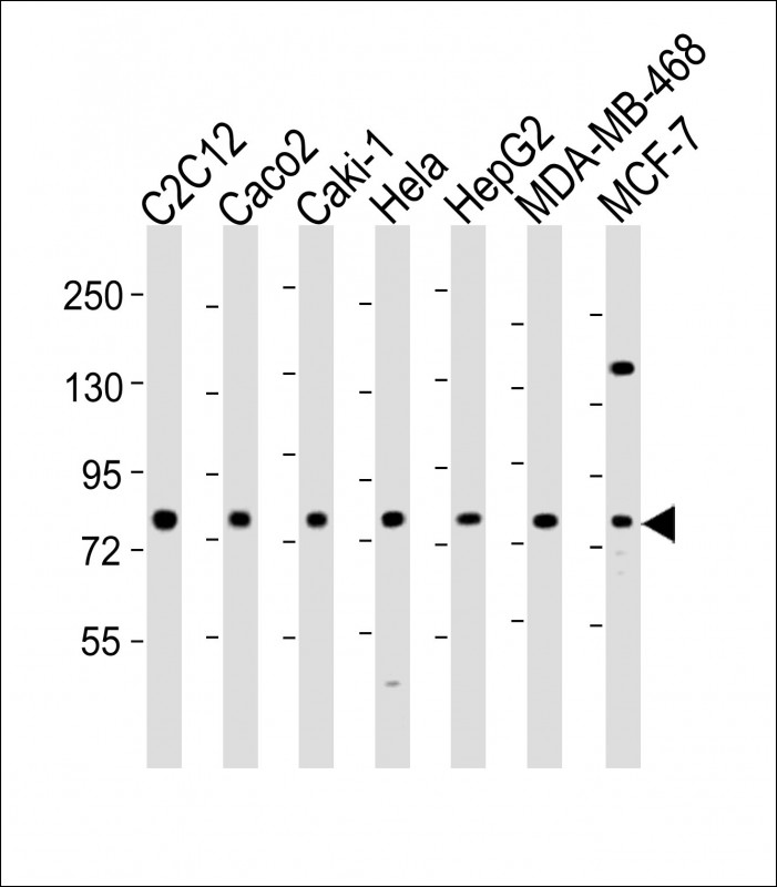
LPP Antibody (N-Term)
Purified Rabbit Polyclonal Antibody (Pab)
- 产品详情
- 实验流程
- 背景知识
Application
| WB, E |
|---|---|
| Primary Accession | Q93052 |
| Other Accession | Q8BFW7, Q5XI07 |
| Reactivity | Human, Mouse |
| Predicted | Mouse, Rat |
| Host | Rabbit |
| Clonality | polyclonal |
| Isotype | Rabbit IgG |
| Calculated MW | 65746 Da |
| Gene ID | 4026 |
|---|---|
| Other Names | Lipoma-preferred partner, LIM domain-containing preferred translocation partner in lipoma, LPP |
| Target/Specificity | This LPP antibody is generated from a rabbit immunized with a KLH conjugated synthetic peptide between 32-66 amino acids from the human LPP. |
| Dilution | WB~~1:2000 E~~Use at an assay dependent concentration. |
| Format | Purified polyclonal antibody supplied in PBS with 0.09% (W/V) sodium azide. This antibody is purified through a protein A column, followed by peptide affinity purification. |
| Storage | Maintain refrigerated at 2-8°C for up to 2 weeks. For long term storage store at -20°C in small aliquots to prevent freeze-thaw cycles. |
| Precautions | LPP Antibody (N-Term) is for research use only and not for use in diagnostic or therapeutic procedures. |
| Name | LPP |
|---|---|
| Function | May play a structural role at sites of cell adhesion in maintaining cell shape and motility. In addition to these structural functions, it may also be implicated in signaling events and activation of gene transcription. May be involved in signal transduction from cell adhesion sites to the nucleus allowing successful integration of signals arising from soluble factors and cell-cell adhesion sites. Also suggested to serve as a scaffold protein upon which distinct protein complexes are assembled in the cytoplasm and in the nucleus. |
| Cellular Location | Nucleus. Cytoplasm. Cell junction. Cell membrane. Note=Found in the nucleus, in the cytoplasm and at cell adhesion sites Shuttles between the cytoplasm and the nucleus. It has been found in sites of cell adhesion such as cell-to-cell contact and focal adhesion which are membrane attachment sites of cells to the extracellular matrix. Mainly nuclear when fused with HMGA2/HMGIC and KMT2A/MLL1 |
| Tissue Location | Expressed in a wide variety of tissues but no or very low expression in brain and peripheral leukocytes |
For Research Use Only. Not For Use In Diagnostic Procedures.
Provided below are standard protocols that you may find useful for product applications.
BACKGROUND
May play a structural role at sites of cell adhesion in maintaining cell shape and motility. In addition to these structural functions, it may also be implicated in signaling events and activation of gene transcription. May be involved in signal transduction from cell adhesion sites to the nucleus allowing successful integration of signals arising from soluble factors and cell-cell adhesion sites. Also suggested to serve as a scaffold protein upon which distinct protein complexes are assembled in the cytoplasm and in the nucleus.
REFERENCES
Petit M.M.R.,et al.Genomics 36:118-129(1996).
Ebert L.,et al.Submitted (JUN-2004) to the EMBL/GenBank/DDBJ databases.
Mural R.J.,et al.Submitted (SEP-2005) to the EMBL/GenBank/DDBJ databases.
Lemke I.,et al.Cytogenet. Cell Genet. 95:153-156(2001).
Petit M.M.,et al.Cancer Genet. Cytogenet. 106:18-23(1998).
终于等到您。ABCEPTA(百远生物)抗体产品。
点击下方“我要评价 ”按钮提交您的反馈信息,您的反馈和评价是我们最宝贵的财富之一,
我们将在1-3个工作日内处理您的反馈信息。
如有疑问,联系:0512-88856768 tech-china@abcepta.com.


