


 癌症的基本特征包括细胞增殖、血管生成、迁移、凋亡逃避机制和细胞永生等。找到癌症发生过程中这些通路的关键标记物和对应的抗体用于检测至关重要。
 为您推荐一个泛素化位点预测神器——泛素化分析工具,可以为您的蛋白的泛素化位点作出预测和评分。
 细胞自噬受体图形绘图工具为你的蛋白的细胞受体结合位点作出预测和评分,识别结合到自噬通路中的蛋白是非常重要的,便于让我们理解自噬在正常生理、病理过程中的作用,如发育、细胞分化、神经退化性疾病、压力条件下、感染和癌症。
NSFL1C Antibody (N-Term)
Purified Rabbit Polyclonal Antibody (Pab)
- 产品详情
- 实验流程
- 背景知识
Application
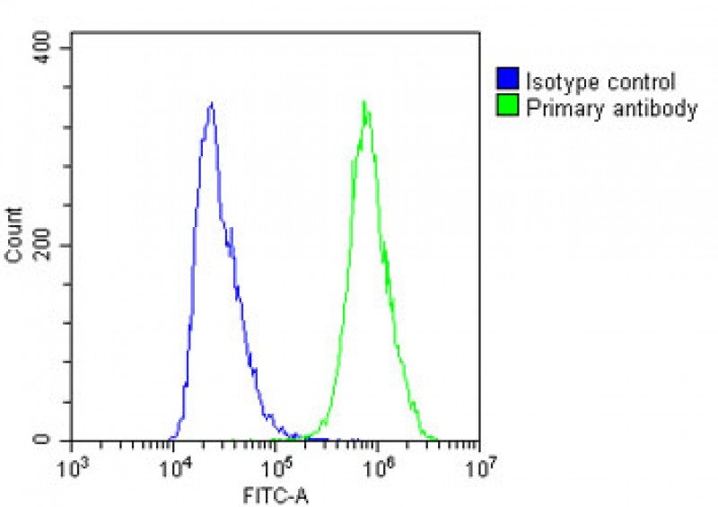
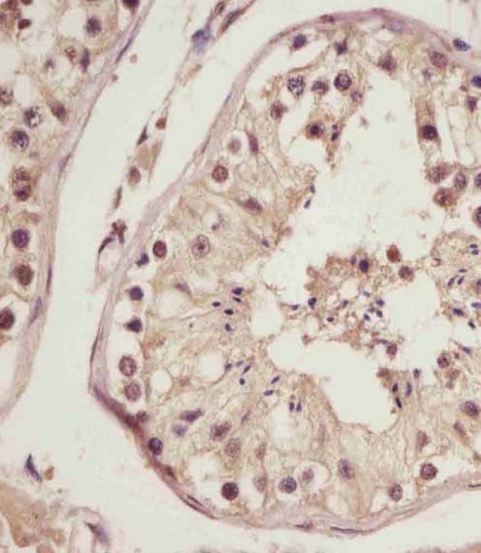
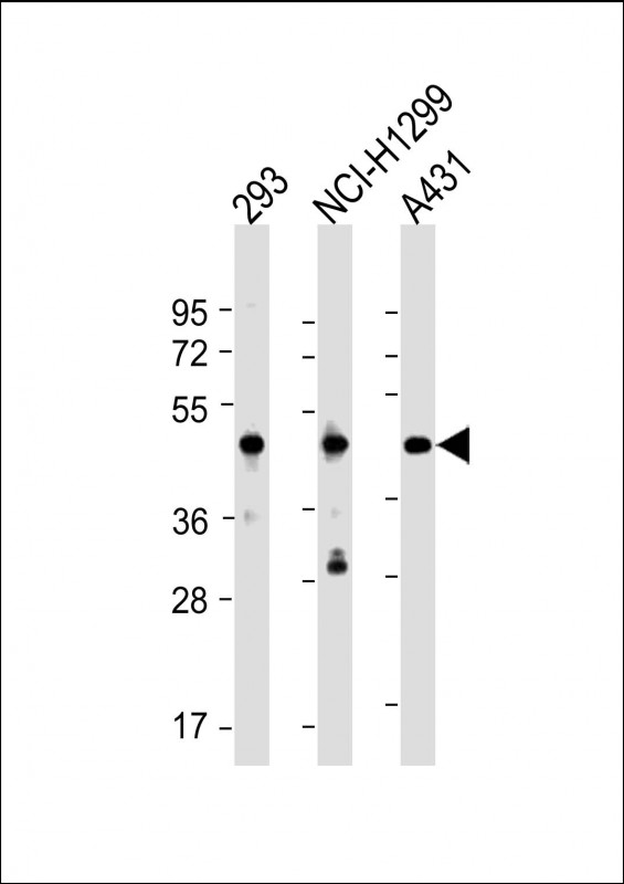
| WB, IHC-P, FC, E |
|---|---|
| Primary Accession | Q9UNZ2 |
| Other Accession | Q3SZC4, Q9CZ44, Q5RBG3, O35987 |
| Reactivity | Human, Rat, Mouse |
| Predicted | Bovine, Mouse, Rat |
| Host | Rabbit |
| Clonality | polyclonal |
| Isotype | Rabbit IgG |
| Calculated MW | 40573 Da |
| Gene ID | 55968 |
|---|---|
| Other Names | NSFL1 cofactor p47, UBX domain-containing protein 2C, p97 cofactor p47, NSFL1C, UBXN2C |
| Target/Specificity | This NSFL1C antibody is generated from a rabbit immunized with a KLH conjugated synthetic peptide between 37-71 amino acids from human NSFL1C. |
| Dilution | WB~~1:2000 IHC-P~~1:100~500 FC~~1:25 E~~Use at an assay dependent concentration. |
| Format | Purified polyclonal antibody supplied in PBS with 0.09% (W/V) sodium azide. This antibody is purified through a protein A column, followed by peptide affinity purification. |
| Storage | Maintain refrigerated at 2-8°C for up to 2 weeks. For long term storage store at -20°C in small aliquots to prevent freeze-thaw cycles. |
| Precautions | NSFL1C Antibody (N-Term) is for research use only and not for use in diagnostic or therapeutic procedures. |
| Name | NSFL1C |
|---|---|
| Synonyms | UBXN2C |
| Function | Reduces the ATPase activity of VCP (By similarity). Necessary for the fragmentation of Golgi stacks during mitosis and for VCP- mediated reassembly of Golgi stacks after mitosis (By similarity). May play a role in VCP-mediated formation of transitional endoplasmic reticulum (tER) (By similarity). Inhibits the activity of CTSL (in vitro) (PubMed:15498563). Together with UBXN2B/p37, regulates the centrosomal levels of kinase AURKA/Aurora A during mitotic progression by promoting AURKA removal from centrosomes in prophase (PubMed:23649807). Also, regulates spindle orientation during mitosis (PubMed:23649807). |
| Cellular Location | Nucleus {ECO:0000250|UniProtKB:O35987}. Golgi apparatus, Golgi stack {ECO:0000250|UniProtKB:O35987}. Chromosome {ECO:0000250|UniProtKB:O35987}. Cytoplasm, cytoskeleton, microtubule organizing center, centrosome {ECO:0000250|UniProtKB:O35987} Note=Predominantly nuclear in interphase cells. Bound to the axial elements of sex chromosomes in pachytene spermatocytes. A small proportion of the protein is cytoplasmic, associated with Golgi stacks Localizes to centrosome during mitotic prophase and metaphase {ECO:0000250|UniProtKB:O35987} |
For Research Use Only. Not For Use In Diagnostic Procedures.
Provided below are standard protocols that you may find useful for product applications.
BACKGROUND
Reduces the ATPase activity of VCP. Necessary for the fragmentation of Golgi stacks during mitosis and for VCP-mediated reassembly of Golgi stacks after mitosis. May play a role in VCP- mediated formation of transitional endoplasmic reticulum (tER) (By similarity). Inhibits the activity of CTSL (in vitro).
REFERENCES
Yue P.,et al.Submitted (AUG-1998) to the EMBL/GenBank/DDBJ databases.
Hu R.-M.,et al.Proc. Natl. Acad. Sci. U.S.A. 97:9543-9548(2000).
Zhang Q.-H.,et al.Genome Res. 10:1546-1560(2000).
Ota T.,et al.Nat. Genet. 36:40-45(2004).
Deloukas P.,et al.Nature 414:865-871(2001).
终于等到您。ABCEPTA(百远生物)抗体产品。
点击下方“我要评价 ”按钮提交您的反馈信息,您的反馈和评价是我们最宝贵的财富之一,
我们将在1-3个工作日内处理您的反馈信息。
如有疑问,联系:0512-88856768 tech-china@abcepta.com.


