


 癌症的基本特征包括细胞增殖、血管生成、迁移、凋亡逃避机制和细胞永生等。找到癌症发生过程中这些通路的关键标记物和对应的抗体用于检测至关重要。
 为您推荐一个泛素化位点预测神器——泛素化分析工具,可以为您的蛋白的泛素化位点作出预测和评分。
 细胞自噬受体图形绘图工具为你的蛋白的细胞受体结合位点作出预测和评分,识别结合到自噬通路中的蛋白是非常重要的,便于让我们理解自噬在正常生理、病理过程中的作用,如发育、细胞分化、神经退化性疾病、压力条件下、感染和癌症。
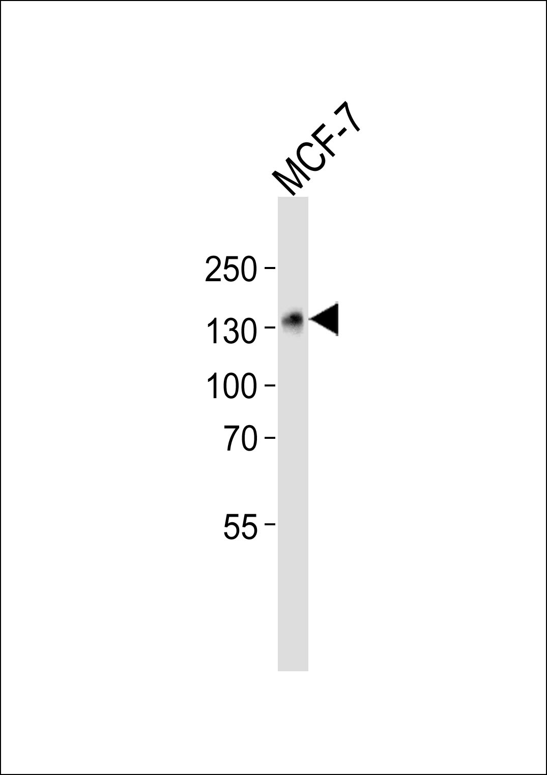
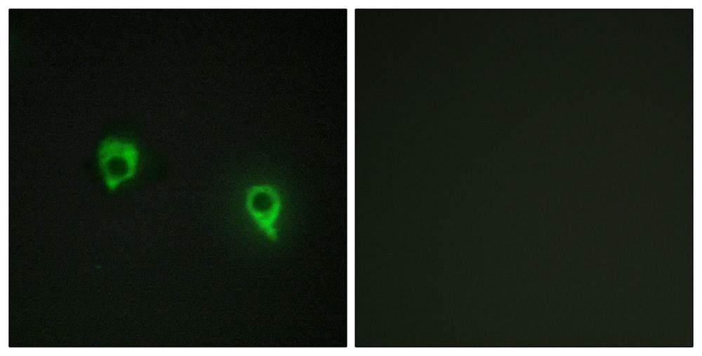
AKAP1 Antibody
Purified Rabbit Polyclonal Antibody (Pab)
- 产品详情
- 实验流程
- 背景知识
Application
| WB, IF |
|---|---|
| Primary Accession | Q92667 |
| Reactivity | Human |
| Host | Rabbit |
| Clonality | polyclonal |
| Calculated MW | 97 KDa |
| Other Names | A-kinase anchor protein 1, mitochondrial, A-kinase anchor protein 149 kDa, AKAP 149, Dual specificity A-kinase-anchoring protein 1, D-AKAP-1, Protein kinase A-anchoring protein 1, PRKA1, Spermatid A-kinase anchor protein 84, S-AKAP84, AKAP1, AKAP149, PRKA1 |
|---|---|
| Dilution | WB~~ 1:1000 IF~~1:100 |
| Format | Rabbit IgG in phosphate buffered saline (without Mg2+ and Ca2+), pH 7.4, 150mM NaCl, 0.09% (W/V) sodium azide and 50% glycerol. |
| Storage Conditions | -20℃ |
For Research Use Only. Not For Use In Diagnostic Procedures.
Provided below are standard protocols that you may find useful for product applications.
BACKGROUND
Binds to type I and II regulatory subunits of protein kinase A and anchors them to the cytoplasmic face of the mitochondrial outer membrane.
REFERENCES
Trendelenburg G.,et al.Biochem. Biophys. Res. Commun. 225:313-319(1996).
Lin R.-Y.,et al.J. Biol. Chem. 270:27804-27811(1995).
Ota T.,et al.Nat. Genet. 36:40-45(2004).
Mural R.J.,et al.Submitted (SEP-2005) to the EMBL/GenBank/DDBJ databases.
Hillman R.T.,et al.Genome Biol. 5:R8.1-R8.16(2004).
终于等到您。ABCEPTA(百远生物)抗体产品。
点击下方“我要评价 ”按钮提交您的反馈信息,您的反馈和评价是我们最宝贵的财富之一,
我们将在1-3个工作日内处理您的反馈信息。
如有疑问,联系:0512-88856768 tech-china@abcepta.com.


