


 癌症的基本特征包括细胞增殖、血管生成、迁移、凋亡逃避机制和细胞永生等。找到癌症发生过程中这些通路的关键标记物和对应的抗体用于检测至关重要。
 为您推荐一个泛素化位点预测神器——泛素化分析工具,可以为您的蛋白的泛素化位点作出预测和评分。
 细胞自噬受体图形绘图工具为你的蛋白的细胞受体结合位点作出预测和评分,识别结合到自噬通路中的蛋白是非常重要的,便于让我们理解自噬在正常生理、病理过程中的作用,如发育、细胞分化、神经退化性疾病、压力条件下、感染和癌症。
beta-Actin Rabbit pAb, Loading Control
beta-Actin Rabbit pAb, Loading Control

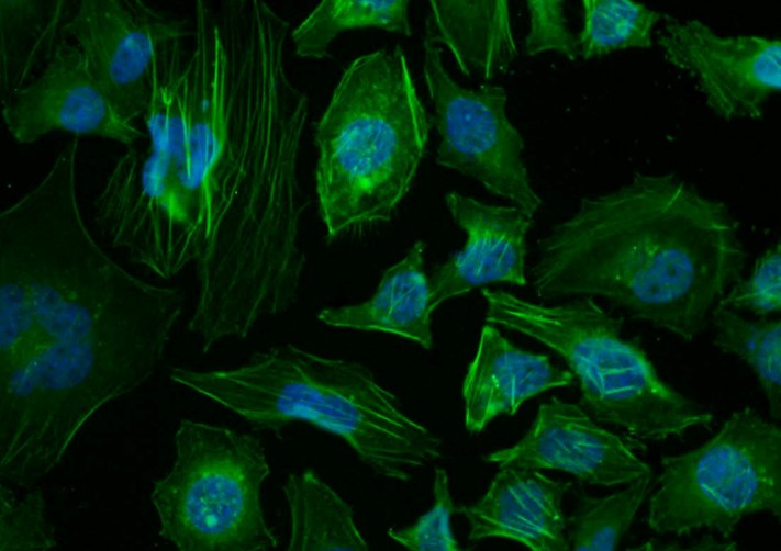

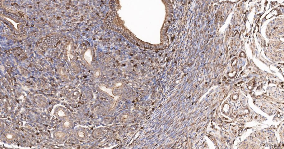
- 产品详情
- 实验流程
- 背景知识
Application 
| WB, IHC-P, IHC-F, IF |
|---|---|
| Primary Accession | P60709 |
| Reactivity | Rat, Human, Mouse |
| Predicted | Pig, Cat, Dog, Bee, Rabbit, Hamster, Chicken, Sheep, Fish |
| Host | Rabbit |
| Clonality | Polyclonal |
| Calculated MW | 41737 Da |
| Physical State | Liquid |
| Immunogen | Synthetic MAP peptide derived from human beta-Actin |
| Epitope Specificity | 1-200/375 |
| Isotype | IgG |
| Purity | affinity purified by Protein A |
| Buffer | 0.01M TBS (pH7.4) with 1% BSA, 0.02% Proclin300 and 50% Glycerol. |
| SUBCELLULAR LOCATION | Cytoplasm, cytoskeleton. |
| DISEASE | Defects in ACTA1 are the cause of nemaline myopathy type 3 (NEM3) [MIM:161800]. A form of nemaline myopathy. Nemaline myopathies are muscular disorders characterized by muscle weakness of varying severity and onset, and abnormal thread-or rod-like structures in muscle fibers on histologic examination. The phenotype at histological level is variable. Some patients present areas devoid of oxidative activity containg (cores) within myofibers. Core lesions are unstructured and poorly circumscribed. Defects in ACTA1 are a cause of myopathy congenital with excess of thin myofilaments (MPCETM) [MIM:161800]. A congenital muscular disorder characterized at histological level by areas of sarcoplasm devoid of normal myofibrils and mitochondria, and replaced with dense masses of thin filaments. Central cores, rods, ragged red fibers, and necrosis are absent. |
| Important Note | This product as supplied is intended for research use only, not for use in human, therapeutic or diagnostic applications. |
| Background Descriptions | Actin is a highly conserved protein and an essential component of cell cytoskeleton and plays an important role in cytoplasmic streaming, cell shape determination, cell division, organelle movement and extension growth. Preferentially expressed in young and expanding tissues, floral organ primordia, developing seeds and emerging inflorescence. Antibodies against plant Actin are useful as loading controls for Western Blotting. |
| Gene ID | 60 |
|---|---|
| Other Names | Actin, cytoplasmic 1, 3.6.4.-, Beta-actin, Actin, cytoplasmic 1, N-terminally processed, ACTB |
| Dilution | WB=1:5000-50000,IHC-P=1:200-1000,IHC-F=1:200-1000,ICC/IF=1:100-500,IF=1:200-1000,Flow-Cyt=1 µg/Test |
| Storage | Store at -20 °C for one year. Avoid repeated freeze/thaw cycles. When reconstituted in sterile pH 7.4 0.01M PBS or diluent of antibody the antibody is stable for at least two weeks at 2-4 °C. |
For Research Use Only. Not For Use In Diagnostic Procedures.
| Name | ACTB |
|---|---|
| Function | Actin is a highly conserved protein that polymerizes to produce filaments that form cross-linked networks in the cytoplasm of cells (PubMed:25255767, PubMed:29581253). Actin exists in both monomeric (G-actin) and polymeric (F-actin) forms, both forms playing key functions, such as cell motility and contraction (PubMed:29581253). In addition to their role in the cytoplasmic cytoskeleton, G- and F- actin also localize in the nucleus, and regulate gene transcription and motility and repair of damaged DNA (PubMed:29925947). Plays a role in the assembly of the gamma-tubulin ring complex (gTuRC), which regulates the minus-end nucleation of alpha-beta tubulin heterodimers that grow into microtubule protafilaments (PubMed:39321809, PubMed:38609661). Part of the ACTR1A/ACTB filament around which the dynactin complex is built (By similarity). The dynactin multiprotein complex activates the molecular motor dynein for ultra-processive transport along microtubules (By similarity). |
| Cellular Location | Cytoplasm, cytoskeleton. Nucleus Note=Localized in cytoplasmic mRNP granules containing untranslated mRNAs. |
Provided below are standard protocols that you may find useful for product applications.
BACKGROUND
This product as supplied is intended for research use only, not for use in human, therapeutic or diagnostic applications.
REFERENCES
Ponte P.,et al.Nucleic Acids Res. 12:1687-1696(1984).
Nakajima-Iijima S.,et al.Proc. Natl. Acad. Sci. U.S.A. 82:6133-6137(1985).
Ohmori H.,et al.J. Cell Biol. 116:933-941(1992).
Hillier L.W.,et al.Nature 424:157-164(2003).
Gevaert K.,et al.Nat. Biotechnol. 21:566-569(2003).

终于等到您。ABCEPTA(百远生物)抗体产品。
点击下方“我要评价 ”按钮提交您的反馈信息,您的反馈和评价是我们最宝贵的财富之一,
我们将在1-3个工作日内处理您的反馈信息。
如有疑问,联系:0512-88856768 tech-china@abcepta.com.


