


 癌症的基本特征包括细胞增殖、血管生成、迁移、凋亡逃避机制和细胞永生等。找到癌症发生过程中这些通路的关键标记物和对应的抗体用于检测至关重要。
 为您推荐一个泛素化位点预测神器——泛素化分析工具,可以为您的蛋白的泛素化位点作出预测和评分。
 细胞自噬受体图形绘图工具为你的蛋白的细胞受体结合位点作出预测和评分,识别结合到自噬通路中的蛋白是非常重要的,便于让我们理解自噬在正常生理、病理过程中的作用,如发育、细胞分化、神经退化性疾病、压力条件下、感染和癌症。
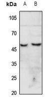
Anti-CHRNA7 Antibody
- 产品详情
- 实验流程
- 背景知识
Application
| WB |
|---|---|
| Primary Accession | P36544 |
| Other Accession | Q494W8 |
| Reactivity | Human, Mouse, Rat |
| Host | Rabbit |
| Clonality | Polyclonal |
| Calculated MW | 56449 Da |
| Gene ID | 1139;89832 |
|---|---|
| Other Names | CHRNA7; NACHRA7; Neuronal acetylcholine receptor subunit alpha-7; CHRFAM7A; CHRNA7-FAM7A fusion protein; CHRNA7-DR1; D-10 |
| Target/Specificity | Recognizes endogenous levels of CHRNA7 protein. |
| Dilution | WB~~1/500 - 1/1000 |
| Format | Liquid in 0.42% Potassium phosphate, 0.87% Sodium chloride, pH 7.3, 30% glycerol, and 0.09% (W/V) sodium azide. |
| Storage | Store at -20 °C.Stable for 12 months from date of receipt |
| Name | CHRNA7 (HGNC:1960) |
|---|---|
| Synonyms | NACHRA7 |
| Function | Component of neuronal acetylcholine receptors (nAChRs) that function as pentameric, ligand-gated cation channels with high calcium permeability among other activities. nAChRs are excitatory neurotrasnmitter receptors formed by a collection of nAChR subunits known to mediate synaptic transmission in the nervous system and the neuromuscular junction. Each nAchR subunit confers differential attributes to channel properties, including activation, deactivation and desensitization kinetics, pH sensitivity, cation permeability, and binding to allosteric modulators (PubMed:15609996, PubMed:33735609, PubMed:8145738). CHRNA7 forms homopentameric neuronal acetylcholine receptors abundantly expressed in the central nervous system, characterized by fast desensitization and high calcium permeability (PubMed:31560909, PubMed:33735609, PubMed:38382524, PubMed:8145738). Also forms heteropentamers with CHRNB2, mainly expressed in basal forebrain cholinergic neurons. Involved in the modulation of calcium- dependent signaling pathways and influences the release of neurotransmitters, including dopamine, glutamate and GABA (PubMed:33239400). Also expressed in non-neuronal cells such as immune cells like lymphocytes, monocytes and macrophages (PubMed:12508119, PubMed:16968406, PubMed:25259522). In T cells, activation induces metabotropic signaling that results in an increase of intracellular Ca2+ concentrations, independent of ionotropic receptor functions (PubMed:17709503). In macrophages, required for acetylcholine-mediated inhibition of TNF and other inflammatory cytokine release (PubMed:12508119). Once activated by acetylcholine, nicotine or other agonists, selectively inhibits production of pro-inflammatory cytokines while leaving anti-inflammatory cytokines undisturbed (PubMed:12508119, PubMed:25259522). Stimulates the cholinergic anti-inflammatory pathway, controlling inflammation by inhibiting NFKB nuclear translocation and activating the JAK2-STAT3 pathway, independently of ion channel activity (PubMed:16968406, PubMed:25259522). Also expressed in the urothelium where it modulates reflex bladder activity by increasing intracellular calcium through internal stores and decreasing basal ATP release (By similarity). |
| Cellular Location | Postsynaptic cell membrane {ECO:0000250|UniProtKB:Q05941}; Multi-pass membrane protein. Cell membrane; Multi-pass membrane protein. Note=TMEM35A/NACHO promotes its trafficking to the cell membrane (PubMed:27789755). RIC3 promotes its trafficking to the cell membrane (By similarity) {ECO:0000250|UniProtKB:Q05941, ECO:0000269|PubMed:27789755} |
| Tissue Location | Expressed in neuronal cells (PubMed:8145738). Expressed in macrophages (at protein level) (PubMed:12508119) |
Research Areas
For Research Use Only. Not For Use In Diagnostic Procedures.
Application Protocols
Provided below are standard protocols that you may find useful for product applications.
BACKGROUND
Rabbit polyclonal antibody to CHRNA7
终于等到您。ABCEPTA(百远生物)抗体产品。
点击下方“我要评价 ”按钮提交您的反馈信息,您的反馈和评价是我们最宝贵的财富之一,
我们将在1-3个工作日内处理您的反馈信息。
如有疑问,联系:0512-88856768 tech-china@abcepta.com.
¥ 1,500.00
Cat# AP53661


