


 癌症的基本特征包括细胞增殖、血管生成、迁移、凋亡逃避机制和细胞永生等。找到癌症发生过程中这些通路的关键标记物和对应的抗体用于检测至关重要。
 为您推荐一个泛素化位点预测神器——泛素化分析工具,可以为您的蛋白的泛素化位点作出预测和评分。
 细胞自噬受体图形绘图工具为你的蛋白的细胞受体结合位点作出预测和评分,识别结合到自噬通路中的蛋白是非常重要的,便于让我们理解自噬在正常生理、病理过程中的作用,如发育、细胞分化、神经退化性疾病、压力条件下、感染和癌症。
Anti-EPHB1/2 Antibody
- 产品详情
- 实验流程
- 背景知识
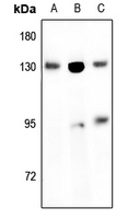
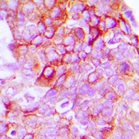
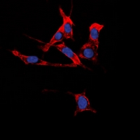
Application
| WB, IF |
|---|---|
| Primary Accession | P54762 |
| Other Accession | P29323 |
| Reactivity | Human, Mouse, Rat |
| Host | Rabbit |
| Clonality | Polyclonal |
| Calculated MW | 109885 Da |
| Gene ID | 2047 |
|---|---|
| Other Names | EPHB1; ELK; EPHT2; HEK6; NET; Ephrin type-B receptor 1; ELK; EPH tyrosine kinase 2; EPH-like kinase 6; EK6; hEK6; Neuronally-expressed EPH-related tyrosine kinase; NET; Tyrosine-protein kinase receptor EPH-2; EPHB2; DRT; EPHT3; EPTH3; ERK; HEK5; TYRO5; Ephrin type-B receptor 2; Developmentally-regulated Eph-related tyrosine kinase; ELK-related tyrosine kinase; EPH tyrosine kinase 3; EPH-like kinase 5; EK5; hEK5; Renal carcinoma antigen NY-REN-47; Tyrosine-protein kinase TYRO5; Tyrosine-protein kinase receptor EPH-3 |
| Target/Specificity | KLH-conjugated synthetic peptide encompassing a sequence within the center region of human EPHB1/2. The exact sequence is proprietary. |
| Dilution | WB~~1/500 - 1/1000 IF~~1/50 - 1/200 |
| Format | Liquid in 0.42% Potassium phosphate, 0.87% Sodium chloride, pH 7.3, 30% glycerol, and 0.09% (W/V) sodium azide. |
| Storage | Store at -20 °C.Stable for 12 months from date of receipt |
| Name | EPHB1 |
|---|---|
| Synonyms | ELK, EPHT2, HEK6, NET |
| Function | Receptor tyrosine kinase which binds promiscuously transmembrane ephrin-B family ligands residing on adjacent cells, leading to contact-dependent bidirectional signaling into neighboring cells. The signaling pathway downstream of the receptor is referred to as forward signaling while the signaling pathway downstream of the ephrin ligand is referred to as reverse signaling. Cognate/functional ephrin ligands for this receptor include EFNB1, EFNB2 and EFNB3. During nervous system development, regulates retinal axon guidance redirecting ipsilaterally ventrotemporal retinal ganglion cells axons at the optic chiasm midline. This probably requires repulsive interaction with EFNB2. In the adult nervous system together with EFNB3, regulates chemotaxis, proliferation and polarity of the hippocampus neural progenitors. In addition to its role in axon guidance also plays an important redundant role with other ephrin-B receptors in development and maturation of dendritic spines and synapse formation. May also regulate angiogenesis. More generally, may play a role in targeted cell migration and adhesion. Upon activation by EFNB1 and probably other ephrin-B ligands activates the MAPK/ERK and the JNK signaling cascades to regulate cell migration and adhesion respectively. Involved in the maintenance of the pool of satellite cells (muscle stem cells) by promoting their self-renewal and reducing their activation and differentiation (By similarity). |
| Cellular Location | Cell membrane; Single-pass type I membrane protein Early endosome membrane. Cell projection, dendrite {ECO:0000250|UniProtKB:Q8CBF3} |
| Tissue Location | Preferentially expressed in brain. |
Research Areas
For Research Use Only. Not For Use In Diagnostic Procedures.
Application Protocols
Provided below are standard protocols that you may find useful for product applications.
BACKGROUND
Rabbit polyclonal antibody to EPHB1/2
终于等到您。ABCEPTA(百远生物)抗体产品。
点击下方“我要评价 ”按钮提交您的反馈信息,您的反馈和评价是我们最宝贵的财富之一,
我们将在1-3个工作日内处理您的反馈信息。
如有疑问,联系:0512-88856768 tech-china@abcepta.com.
¥ 1,500.00
Cat# AP54131


