


 癌症的基本特征包括细胞增殖、血管生成、迁移、凋亡逃避机制和细胞永生等。找到癌症发生过程中这些通路的关键标记物和对应的抗体用于检测至关重要。
 为您推荐一个泛素化位点预测神器——泛素化分析工具,可以为您的蛋白的泛素化位点作出预测和评分。
 细胞自噬受体图形绘图工具为你的蛋白的细胞受体结合位点作出预测和评分,识别结合到自噬通路中的蛋白是非常重要的,便于让我们理解自噬在正常生理、病理过程中的作用,如发育、细胞分化、神经退化性疾病、压力条件下、感染和癌症。
PTPRS Rabbit pAb
PTPRS Rabbit pAb
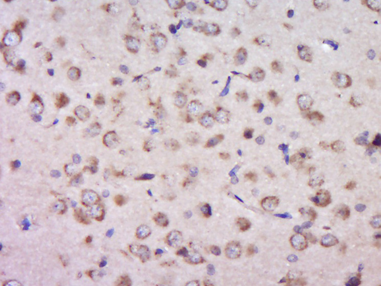
- 产品详情
- 实验流程
- 背景知识
Application 
| IHC-P, IHC-F, IF |
|---|---|
| Primary Accession | Q13332 |
| Reactivity | Rat |
| Predicted | Human, Mouse |
| Host | Rabbit |
| Clonality | Polyclonal |
| Calculated MW | 217041 Da |
| Physical State | Liquid |
| Immunogen | KLH conjugated synthetic peptide derived from human PTPRS |
| Epitope Specificity | 1001-1100/1948 |
| Isotype | IgG |
| Purity | affinity purified by Protein A |
| Buffer | Preservative: 0.02% Proclin300, Constituents: 1% BSA, 0.01M PBS, pH7.4. |
| SUBCELLULAR LOCATION | Membrane; Single-pass type I membrane protein. |
| Important Note | This product as supplied is intended for research use only, not for use in human, therapeutic or diagnostic applications. |
| Background Descriptions | Protein tyrosine phosphatases, or PTPs, are type I transmembrane proteins, membrane associated proteins or proteins localized in nuclei. Examples of transmembrane PTPs are LAR, PTP alpha, PTP beta, PTP gamma, PTP delta, PTP epsilon, PTP zeta, PTP thet, PTP upsilon and PTPs. Transmembrane PTPsigma play diverse roles during development and in adult tissues. Immunodepletion studies have suggested LAR to be a regulator of insulin receptor phosphorylation. PTP alpha activity is increased twofold in response to phorbol ester stimulation, resulting in serine phosphorylation either directly or indirectly by members of the PKC family. Overexpression of v-H-Ras and Neu, but not Myc or Int2, in mammary tumors has been shown to induce PTPe expression. An alternative splicing event leads to a nervous tissue-specific chondroitin sulfate proteoglycan called phosphacan, which represents the amino terminal portion of PTP omega. PTP thet and PTP?share a conserved amino terminal 160 amino acid MAM domain which facilitates homophilic binding. PTP upsilon localizes to points of cell contact and may be involved in regulating the assembly and disassembly of cadherin/catenin complexes in vivo. PTPsigma contains an extracellular region, a single transmembrane segment and two tandem intracytoplasmic catalytic domains, and thus represents a receptor-type PTP. PTPsigma may also be involved in the molecular control of adult nerve repair. Four alternatively spliced transcript variants, which encode distinct proteins, have been reported. |
| Gene ID | 5802 |
|---|---|
| Other Names | Receptor-type tyrosine-protein phosphatase S, R-PTP-S, 3.1.3.48, Receptor-type tyrosine-protein phosphatase sigma, R-PTP-sigma, PTPRS |
| Dilution | IHC-P=1:100-500,IHC-F=1:100-500,IF=1:100-500 |
| Storage | Store at -20 °C for one year. Avoid repeated freeze/thaw cycles. When reconstituted in sterile pH 7.4 0.01M PBS or diluent of antibody the antibody is stable for at least two weeks at 2-4 °C. |
For Research Use Only. Not For Use In Diagnostic Procedures.
| Name | PTPRS |
|---|---|
| Function | Cell surface receptor that binds to glycosaminoglycans, including chondroitin sulfate proteoglycans and heparan sulfate proteoglycan (PubMed:21454754). Binding to chondroitin sulfate and heparan sulfate proteoglycans has opposite effects on PTPRS oligomerization and regulation of neurite outgrowth. Contributes to the inhibition of neurite and axonal outgrowth by chondroitin sulfate proteoglycans, also after nerve transection. Plays a role in stimulating neurite outgrowth in response to the heparan sulfate proteoglycan GPC2. Required for normal brain development, especially for normal development of the pituitary gland and the olfactory bulb. Functions as a tyrosine phosphatase (PubMed:8524829). Mediates dephosphorylation of NTRK1, NTRK2 and NTRK3 (By similarity). Plays a role in down-regulation of signaling cascades that lead to the activation of Akt and MAP kinases (By similarity). Down-regulates TLR9- mediated activation of NF-kappa-B, as well as production of TNF, interferon alpha and interferon beta (PubMed:26231120). |
| Cellular Location | Cell membrane; Single-pass type I membrane protein. Cell projection, axon {ECO:0000250|UniProtKB:B0V2N1}. Perikaryon {ECO:0000250|UniProtKB:B0V2N1}. Cytoplasmic vesicle, secretory vesicle, synaptic vesicle membrane {ECO:0000250|UniProtKB:Q64605}. Synapse, synaptosome {ECO:0000250|UniProtKB:Q64605}. Postsynaptic density {ECO:0000250|UniProtKB:Q64605}. Cell projection, neuron projection {ECO:0000250|UniProtKB:B0V2N1}. Cell projection, growth cone {ECO:0000250|UniProtKB:B0V2N1}. Note=Is rapidly internalized when dendritic cells are stimulated with the TLR9 ligand cytidine-phosphate- guanosine (CpG) (PubMed:26231120). Detected in a punctate pattern along neurites and axon growth cones (By similarity) {ECO:0000250|UniProtKB:B0V2N1, ECO:0000269|PubMed:26231120} |
| Tissue Location | Detected in peripheral blood plasmacytoid dendritic cells (at protein level) (PubMed:26231120). Detected in all tissues tested except for placenta and liver (PubMed:8524829, PubMed:8992885) Detected in peripheral blood plasmacytoid dendritic cells (PubMed:26231120). |
Research Areas
Application Protocols
Provided below are standard protocols that you may find useful for product applications.
BACKGROUND
This product as supplied is intended for research use only, not for use in human, therapeutic or diagnostic applications.

终于等到您。ABCEPTA(百远生物)抗体产品。
点击下方“我要评价 ”按钮提交您的反馈信息,您的反馈和评价是我们最宝贵的财富之一,
我们将在1-3个工作日内处理您的反馈信息。
如有疑问,联系:0512-88856768 tech-china@abcepta.com.


