


 癌症的基本特征包括细胞增殖、血管生成、迁移、凋亡逃避机制和细胞永生等。找到癌症发生过程中这些通路的关键标记物和对应的抗体用于检测至关重要。
 为您推荐一个泛素化位点预测神器——泛素化分析工具,可以为您的蛋白的泛素化位点作出预测和评分。
 细胞自噬受体图形绘图工具为你的蛋白的细胞受体结合位点作出预测和评分,识别结合到自噬通路中的蛋白是非常重要的,便于让我们理解自噬在正常生理、病理过程中的作用,如发育、细胞分化、神经退化性疾病、压力条件下、感染和癌症。
WWP1 Polyclonal Antibody
Purified Rabbit Polyclonal Antibody (Pab)
- 产品详情
- 实验流程
Application
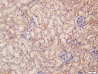
| IHC-P, IHC-F, IF, E |
|---|---|
| Primary Accession | Q9H0M0 |
| Reactivity | Rat, Dog, Bovine |
| Host | Rabbit |
| Clonality | Polyclonal |
| Calculated MW | 105202 Da |
| Physical State | Liquid |
| Immunogen | KLH conjugated synthetic peptide derived from human WWP1 |
| Epitope Specificity | 21-120/922 |
| Isotype | IgG |
| Purity | affinity purified by Protein A |
| Buffer | 0.01M TBS (pH7.4) with 1% BSA, 0.02% Proclin300 and 50% Glycerol. |
| SUBCELLULAR LOCATION | Cytoplasm. Cell membrane; Peripheral membrane protein. Nucleus. |
| SIMILARITY | Contains 1 C2 domain.Contains 1 HECT (E6AP-type E3 ubiquitin-protein ligase) domain. Contains 4 WW domains. |
| SUBUNIT | Binds KLF2 AND HIVEP3. Binds SCNN1A, SCNN1B, SCNN1G, WBP1, WBP2, DRPLA and adenovirus type 2 PIII. Interacts with RNF11. Interacts with SPG20. Interacts with ERBB4 isoforms JM-B CYT-1 and JM-A CYT-1. Interacts with SMAD1, SMAD2, SMAD3, SMAD5, SMAD6, SMAD7, TGFBR1 AND TGFBR2. Associates with the TGFBR1:TGFBR2 receptor complex in presence of SMAD7. Interacts with SKIL isoform 1. Interacts with TP63 isoform 1 and isoform 2. Interacts with STAMBP and RNF11. Interacts with NDFIP1 and NDFIP2 (Probable); this interaction activates the E3 ubiquitin-protein ligase. Interacts with TGIF. |
| Post-translational modifications | Auto-ubiquitinated and ubiquitinated by RNF11. |
| Important Note | This product as supplied is intended for research use only, not for use in human, therapeutic or diagnostic applications. |
| Background Descriptions | WWP1 is an E3 ubiquitin ligase and belongs to a family of NEDD4-like proteins. WWP1 contains 4 tandem WW domains and a HECT (homologous to the E6-associated protein carboxyl terminus) domain. WW domain-containing proteins are found in all eukaryotes and play an important role in the regulation of a wide variety of cellular functions such as protein degradation, transcription, and RNA splicing. The HECT domain of WWP1 has been implicated in regulating the localization and stability of p53 – inhibition of WWP1 results in a decrease in p53 expression, whilst WWP1 mediated stabilization of p53 appears to be associated with an accumulation of cytoplasmic p53. WWP1 also negatively regulates the TGF beta tumor suppressor pathway by inactivating its molecular components (SMAD2, SMAD4 and TGFbetaR1). WWP1 has been implicated in both breast and prostate cancers. |
| Gene ID | 11059 |
|---|---|
| Other Names | NEDD4-like E3 ubiquitin-protein ligase WWP1, 2.3.2.26, Atrophin-1-interacting protein 5, AIP5, HECT-type E3 ubiquitin transferase WWP1, TGIF-interacting ubiquitin ligase 1, Tiul1, WW domain-containing protein 1, WWP1 |
| Target/Specificity | Detected in heart, placenta, pancreas, kidney, liver, skeletal muscle, bone marrow, fetal brain, and at much lower levels in adult brain and lung. Isoform 1 and isoform 5 predominate in all tissues tested, except in testis and bone marrow, where isoform 5 is expressed at much higher levels than isoform 1. |
| Dilution | IHC-P=1:100-500,IHC-F=1:100-500,IF=1:100-500,ELISA=1:5000-10000 |
| Storage | Store at -20 °C for one year. Avoid repeated freeze/thaw cycles. When reconstituted in sterile pH 7.4 0.01M PBS or diluent of antibody the antibody is stable for at least two weeks at 2-4 °C. |
| Name | WWP1 |
|---|---|
| Function | E3 ubiquitin-protein ligase which accepts ubiquitin from an E2 ubiquitin-conjugating enzyme in the form of a thioester and then directly transfers the ubiquitin to targeted substrates. Ubiquitinates ERBB4 isoforms JM-A CYT-1 and JM-B CYT-1, KLF2, KLF5 and TP63 and promotes their proteasomal degradation. Ubiquitinates RNF11 without targeting it for degradation. Ubiquitinates and promotes degradation of TGFBR1; the ubiquitination is enhanced by SMAD7. Ubiquitinates SMAD6 and SMAD7. Ubiquitinates and promotes degradation of SMAD2 in response to TGF-beta signaling, which requires interaction with TGIF. Activates the Hippo signaling pathway in response to cell contact inhibition and recruitment to the Crumbs complex at the cell membrane (PubMed:34404733). Monoubiquitinates AMOTL2 which facilitates its interaction with and activation of LATS2 (PubMed:34404733). LATS2 then phosphorylates YAP1, excluding it from the nucleus and therefore ultimately represses YAP1-driven transcription of target genes (PubMed:34404733). |
| Cellular Location | Cytoplasm. Cell membrane; Peripheral membrane protein {ECO:0000250|UniProtKB:Q8BZZ3}. Nucleus {ECO:0000250|UniProtKB:Q8BZZ3} Cell junction. Note=Translocates to the plasma membrane in response to increased cell-cell contact inhibition and subsequent interaction with the Crumbs complex |
| Tissue Location | Detected in heart, placenta, pancreas, kidney, liver, skeletal muscle, bone marrow, fetal brain, and at much lower levels in adult brain and lung. Isoform 1 and isoform 5 predominate in all tissues tested, except in testis and bone marrow, where isoform 5 is expressed at much higher levels than isoform 1 |
Research Areas
For Research Use Only. Not For Use In Diagnostic Procedures.
Application Protocols
Provided below are standard protocols that you may find useful for product applications.
终于等到您。ABCEPTA(百远生物)抗体产品。
点击下方“我要评价 ”按钮提交您的反馈信息,您的反馈和评价是我们最宝贵的财富之一,
我们将在1-3个工作日内处理您的反馈信息。
如有疑问,联系:0512-88856768 tech-china@abcepta.com.
¥ 1,500.00
Cat# AP58505


