


 癌症的基本特征包括细胞增殖、血管生成、迁移、凋亡逃避机制和细胞永生等。找到癌症发生过程中这些通路的关键标记物和对应的抗体用于检测至关重要。
 为您推荐一个泛素化位点预测神器——泛素化分析工具,可以为您的蛋白的泛素化位点作出预测和评分。
 细胞自噬受体图形绘图工具为你的蛋白的细胞受体结合位点作出预测和评分,识别结合到自噬通路中的蛋白是非常重要的,便于让我们理解自噬在正常生理、病理过程中的作用,如发育、细胞分化、神经退化性疾病、压力条件下、感染和癌症。
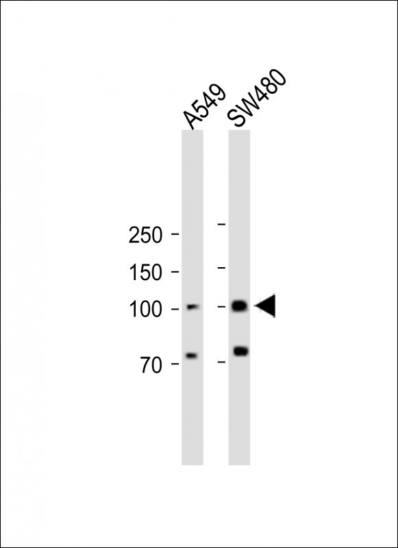
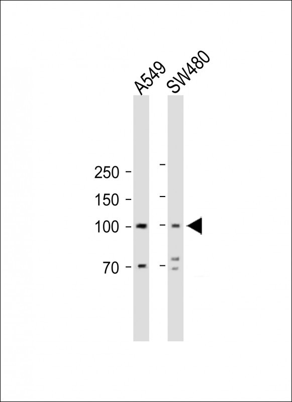
RASGRP1 Antibody (Center)
Affinity Purified Rabbit Polyclonal Antibody (Pab)
- 产品详情
- 文献引用 : 1
- 实验流程
Application
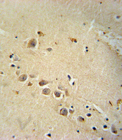
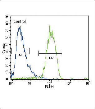
| WB, IHC-P, FC, E |
|---|---|
| Primary Accession | O95267 |
| Other Accession | Q9R1K8, Q9Z1S3, NP_005730.2, NP_001122074.1 |
| Reactivity | Human |
| Predicted | Mouse, Rat |
| Host | Rabbit |
| Clonality | Polyclonal |
| Isotype | Rabbit IgG |
| Calculated MW | 90402 Da |
| Antigen Region | 495-521 aa |
| Gene ID | 10125 |
|---|---|
| Other Names | RAS guanyl-releasing protein 1, Calcium and DAG-regulated guanine nucleotide exchange factor II, CalDAG-GEFII, Ras guanyl-releasing protein, RASGRP1, RASGRP |
| Target/Specificity | This RASGRP1 antibody is generated from rabbits immunized with a KLH conjugated synthetic peptide between 495-521 amino acids from the Central region of human RASGRP1. |
| Dilution | WB~~1:1000 IHC-P~~1:100~500 FC~~1:10~50 E~~Use at an assay dependent concentration. |
| Format | Purified polyclonal antibody supplied in PBS with 0.05% (V/V) Proclin 300. This antibody is prepared by Saturated Ammonium Sulfate (SAS) precipitation followed by dialysis against PBS. |
| Storage | Maintain refrigerated at 2-8°C for up to 2 weeks. For long term storage store at -20°C in small aliquots to prevent freeze-thaw cycles. |
| Precautions | RASGRP1 Antibody (Center) is for research use only and not for use in diagnostic or therapeutic procedures. |
| Name | RASGRP1 |
|---|---|
| Synonyms | RASGRP |
| Function | Functions as a calcium- and diacylglycerol (DAG)-regulated nucleotide exchange factor specifically activating Ras through the exchange of bound GDP for GTP (PubMed:15899849, PubMed:23908768, PubMed:27776107, PubMed:29155103). Activates the Erk/MAP kinase cascade (PubMed:15899849). Regulates T-cell/B-cell development, homeostasis and differentiation by coupling T-lymphocyte/B-lymphocyte antigen receptors to Ras (PubMed:10807788, PubMed:12839994, PubMed:27776107, PubMed:29155103). Regulates NK cell cytotoxicity and ITAM-dependent cytokine production by activation of Ras-mediated ERK and JNK pathways (PubMed:19933860). Functions in mast cell degranulation and cytokine secretion, regulating FcERI-evoked allergic responses. May also function in differentiation of other cell types (PubMed:12845332). |
| Cellular Location | Cytoplasm, cytosol. Cell membrane; Peripheral membrane protein. Golgi apparatus membrane; Peripheral membrane protein. Endoplasmic reticulum membrane; Peripheral membrane protein Note=Found both in the cytosol and associated with membranes Relocalization to the cell membrane upon activation is F-actin- dependent. Translocates to the Golgi in response to phorbol ester or nerve growth factor. Localizes to somata and dendrites but not to axons of hippocampal pyramidal cells (By similarity). |
| Tissue Location | Expressed in brain with higher expression in cerebellum, cerebral cortex and amygdala. Expressed in the hematopoietic system. Expressed in T-cells (at protein level) Expressed in NK cells (at protein level) (PubMed:19933860) |
Research Areas
For Research Use Only. Not For Use In Diagnostic Procedures.

Application Protocols
Provided below are standard protocols that you may find useful for product applications.
终于等到您。ABCEPTA(百远生物)抗体产品。
点击下方“我要评价 ”按钮提交您的反馈信息,您的反馈和评价是我们最宝贵的财富之一,
我们将在1-3个工作日内处理您的反馈信息。
如有疑问,联系:0512-88856768 tech-china@abcepta.com.
¥ 699.00
Cat# AP5856C


