


 癌症的基本特征包括细胞增殖、血管生成、迁移、凋亡逃避机制和细胞永生等。找到癌症发生过程中这些通路的关键标记物和对应的抗体用于检测至关重要。
 为您推荐一个泛素化位点预测神器——泛素化分析工具,可以为您的蛋白的泛素化位点作出预测和评分。
 细胞自噬受体图形绘图工具为你的蛋白的细胞受体结合位点作出预测和评分,识别结合到自噬通路中的蛋白是非常重要的,便于让我们理解自噬在正常生理、病理过程中的作用,如发育、细胞分化、神经退化性疾病、压力条件下、感染和癌症。
TRPV5 Rabbit pAb
TRPV5 Rabbit pAb
- 产品详情
- 实验流程
- 背景知识
| Primary Accession | Q9NQA5 |
|---|---|
| Reactivity | Human |
| Predicted | Mouse, Rat, Dog, Pig, Horse, Rabbit, Sheep |
| Host | Rabbit |
| Clonality | Polyclonal |
| Calculated MW | 82562 Da |
| Physical State | Liquid |
| Immunogen | KLH conjugated synthetic peptide derived from human TRPV5 |
| Epitope Specificity | 201-300/729 |
| Isotype | IgG |
| Purity | affinity purified by Protein A |
| Buffer | 0.01M TBS (pH7.4) with 1% BSA, 0.02% Proclin300 and 50% Glycerol. |
| SUBCELLULAR LOCATION | Apical cell membrane. Colocalized with S100A10 and ANAX2 along the apical domain of kidney distal tubular cells (By similarity). The expression of the glycosylated form in the cell membrane is increased in the presence of WNK3. |
| SIMILARITY | Belongs to the transient receptor (TC 1.A.4) family. TrpV subfamily. TRPV5 sub-subfamily.Contains 5 ANK repeats. |
| SUBUNIT | Homotetramer and probably heterotetramer with TRPV6. Interacts with TRPV6. Interacts with S100A10 and probably with the ANAX2-S100A10 heterotetramer. The interaction with S100A10 is required for the trafficking to the plasma membrane. Interacts with calmodulin. Interacts with BSPRY, which results in its inactivation (By similarity). |
| Post-translational modifications | Glycosylated. |
| Important Note | This product as supplied is intended for research use only, not for use in human, therapeutic or diagnostic applications. |
| Background Descriptions | Transient receptor potential (TRP) proteins are cation-sensitive channels that modulate a myriad of cellular functions, including temperature sensation and vasoregulation Transcribed from a gene adjacent to VR-1, the thermal-sensitive, capsaicin-insensitive TRPV3 is expressed at warm temperatures; expression increases in response to noxious temperatures. Human TRPV3 is expressed in skin, tongue, dorsal root ganglion, trigeminal ganglion, spinal cord and brain. In addition, TRPV3 is co-expressed in dosal root ganglion neurons with VR-1. TRPV3 associates with VR-1 and may modulate VR-1 activity. The 729 amino acid TRPV5 (ECAC1) protein comprises six transmembrane domains, multiple potential phosphorylation sites, an N-linked glycosylation site and three ankyrin repeat regions. It is abundantly expressed in kidney, jejunum and pancreas, and at lower levels in testis, prostate, placenta, brain, colon and rectum. TRPV5 controls the rate-limiting step of vitamin D3-regulated Ca2+ reabsorption in kidney and intestine; the 5’-flanking region of TRPV5 contains four putative vitamin D3-responsive elements. |
| Gene ID | 56302 |
|---|---|
| Other Names | Transient receptor potential cation channel subfamily V member 5, TrpV5, Calcium transport protein 2, CaT2, Epithelial calcium channel 1, ECaC, ECaC1, Osm-9-like TRP channel 3, OTRPC3, TRPV5, ECAC1 {ECO:0000303|PubMed:10945469} |
| Target/Specificity | Expressed at high levels in kidney, small intestine and pancreas, and at lower levels in testis, prostate, placenta, brain, colon and rectum. |
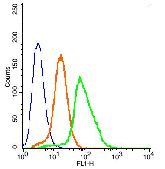
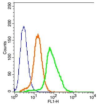
| Dilution | Flow-Cyt=1 µg/Test |
| Storage | Store at -20 °C for one year. Avoid repeated freeze/thaw cycles. When reconstituted in sterile pH 7.4 0.01M PBS or diluent of antibody the antibody is stable for at least two weeks at 2-4 °C. |
| Name | TRPV5 |
|---|---|
| Synonyms | ECAC1 {ECO:0000303|PubMed:10945469} |
| Function | Constitutively active calcium selective cation channel thought to be involved in Ca(2+) reabsorption in kidney and intestine (PubMed:11549322, PubMed:18768590). Required for normal Ca(2+) reabsorption in the kidney distal convoluted tubules (By similarity). The channel is activated by low internal calcium level and the current exhibits an inward rectification (PubMed:11549322, PubMed:18768590). A Ca(2+)-dependent feedback regulation includes fast channel inactivation and slow current decay (By similarity). Heteromeric assembly with TRPV6 seems to modify channel properties. TRPV5-TRPV6 heteromultimeric concatemers exhibit voltage-dependent gating (By similarity). |
| Cellular Location | Apical cell membrane; Multi-pass membrane protein. Note=Colocalized with S100A10 and ANAX2 along the apical domain of kidney distal tubular cells (By similarity) The expression of the glycosylated form in the cell membrane is increased in the presence of WNK3 (PubMed:18768590) {ECO:0000250|UniProtKB:P69744, ECO:0000269|PubMed:18768590} |
| Tissue Location | Expressed at high levels in kidney, small intestine and pancreas, and at lower levels in testis, prostate, placenta, brain, colon and rectum. |
For Research Use Only. Not For Use In Diagnostic Procedures.
Provided below are standard protocols that you may find useful for product applications.
BACKGROUND
Transient receptor potential (TRP) proteins are cation-sensitive channels that modulate a myriad of cellular functions, including temperature sensation and vasoregulation Transcribed from a gene adjacent to VR-1, the thermal-sensitive, capsaicin-insensitive TRPV3 is expressed at warm temperatures; expression increases in response to noxious temperatures. Human TRPV3 is expressed in skin, tongue, dorsal root ganglion, trigeminal ganglion, spinal cord and brain. In addition, TRPV3 is co-expressed in dosal root ganglion neurons with VR-1. TRPV3 associates with VR-1 and may modulate VR-1 activity. The 729 amino acid TRPV5 (ECAC1) protein comprises six transmembrane domains, multiple potential phosphorylation sites, an N-linked glycosylation site and three ankyrin repeat regions. It is abundantly expressed in kidney, jejunum and pancreas, and at lower levels in testis, prostate, placenta, brain, colon and rectum. TRPV5 controls the rate-limiting step of vitamin D3-regulated Ca2+ reabsorption in kidney and intestine; the 5’-flanking region of TRPV5 contains four putative vitamin D3-responsive elements.
终于等到您。ABCEPTA(百远生物)抗体产品。
点击下方“我要评价 ”按钮提交您的反馈信息,您的反馈和评价是我们最宝贵的财富之一,
我们将在1-3个工作日内处理您的反馈信息。
如有疑问,联系:0512-88856768 tech-china@abcepta.com.


