


 癌症的基本特征包括细胞增殖、血管生成、迁移、凋亡逃避机制和细胞永生等。找到癌症发生过程中这些通路的关键标记物和对应的抗体用于检测至关重要。
 为您推荐一个泛素化位点预测神器——泛素化分析工具,可以为您的蛋白的泛素化位点作出预测和评分。
 细胞自噬受体图形绘图工具为你的蛋白的细胞受体结合位点作出预测和评分,识别结合到自噬通路中的蛋白是非常重要的,便于让我们理解自噬在正常生理、病理过程中的作用,如发育、细胞分化、神经退化性疾病、压力条件下、感染和癌症。
Anti-FAP alpha Antibody
Rabbit polyclonal antibody to FAP alpha
- 产品详情
- 实验流程
- 背景知识
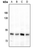
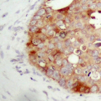
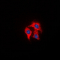
Application
| WB, IF/IC, IHC |
|---|---|
| Primary Accession | Q12884 |
| Other Accession | P97321 |
| Reactivity | Human, Mouse, Rat, Bovine |
| Host | Rabbit |
| Clonality | Polyclonal |
| Calculated MW | 87713 Da |
| Gene ID | 2191 |
|---|---|
| Other Names | Seprase; 170 kDa melanoma membrane-bound gelatinase; Fibroblast activation protein alpha; Integral membrane serine protease |
| Target/Specificity | KLH-conjugated synthetic peptide encompassing a sequence within the center region of human FAP alpha. The exact sequence is proprietary. |
| Dilution | WB~~WB (1/500 - 1/1000), IHC (1/100 - 1/200), IF/IC (1/100 - 1/500) IF/IC~~N/A IHC~~WB (1/500 - 1/1000), IHC (1/100 - 1/200), IF/IC (1/100 - 1/500) |
| Format | Liquid in 0.42% Potassium phosphate, 0.87% Sodium chloride, pH 7.3, 30% glycerol, and 0.09% (W/V) sodium azide. |
| Storage | Store at -20 °C.Stable for 12 months from date of receipt |
| Name | FAP (HGNC:3590) |
|---|---|
| Function | Cell surface glycoprotein serine protease that participates in extracellular matrix degradation and involved in many cellular processes including tissue remodeling, fibrosis, wound healing, inflammation and tumor growth. Both plasma membrane and soluble forms exhibit post-proline cleaving endopeptidase activity, with a marked preference for Ala/Ser-Gly-Pro-Ser/Asn/Ala consensus sequences, on substrate such as alpha-2-antiplasmin SERPINF2 and SPRY2 (PubMed:14751930, PubMed:16223769, PubMed:16410248, PubMed:16480718, PubMed:17381073, PubMed:18095711, PubMed:21288888, PubMed:24371721). Degrade also gelatin, heat-denatured type I collagen, but not native collagen type I and IV, vitronectin, tenascin, laminin, fibronectin, fibrin or casein (PubMed:10347120, PubMed:10455171, PubMed:12376466, PubMed:16223769, PubMed:16651416, PubMed:18095711, PubMed:2172980, PubMed:7923219, PubMed:9065413). Also has dipeptidyl peptidase activity, exhibiting the ability to hydrolyze the prolyl bond two residues from the N-terminus of synthetic dipeptide substrates provided that the penultimate residue is proline, with a preference for Ala-Pro, Ile-Pro, Gly-Pro, Arg-Pro and Pro-Pro (PubMed:10347120, PubMed:10593948, PubMed:16175601, PubMed:16223769, PubMed:16410248, PubMed:16651416, PubMed:17381073, PubMed:21314817, PubMed:24371721, PubMed:24717288). Natural neuropeptide hormones for dipeptidyl peptidase are the neuropeptide Y (NPY), peptide YY (PYY), substance P (TAC1) and brain natriuretic peptide 32 (NPPB) (PubMed:21314817). The plasma membrane form, in association with either DPP4, PLAUR or integrins, is involved in the pericellular proteolysis of the extracellular matrix (ECM), and hence promotes cell adhesion, migration and invasion through the ECM. Plays a role in tissue remodeling during development and wound healing. Participates in the cell invasiveness towards the ECM in malignant melanoma cancers. Enhances tumor growth progression by increasing angiogenesis, collagen fiber degradation and apoptosis and by reducing antitumor response of the immune system. Promotes glioma cell invasion through the brain parenchyma by degrading the proteoglycan brevican. Acts as a tumor suppressor in melanocytic cells through regulation of cell proliferation and survival in a serine protease activity-independent manner. |
| Cellular Location | [Prolyl endopeptidase FAP]: Cell surface. Cell membrane; Single-pass type II membrane protein. Cell projection, lamellipodium membrane; Single-pass type II membrane protein. Cell projection, invadopodium membrane; Single-pass type II membrane protein. Cell projection, ruffle membrane; Single-pass type II membrane protein. Membrane; Single- pass type II membrane protein. Note=Localized on cell surface with lamellipodia and invadopodia membranes and on shed vesicles. Colocalized with DPP4 at invadopodia and lamellipodia membranes of migratory activated endothelial cells in collagenous matrix. Colocalized with DPP4 on endothelial cells of capillary-like microvessels but not large vessels within invasive breast ductal carcinoma. Anchored and enriched preferentially by integrin alpha- 3/beta-1 at invadopodia, plasma membrane protrusions that correspond to sites of cell invasion, in a collagen-dependent manner. Localized at plasma and ruffle membranes in a collagen-independent manner Colocalized with PLAUR preferentially at the cell surface of invadopodia membranes in a cytoskeleton-, integrin- and vitronectin- dependent manner. Concentrated at invadopodia membranes, specialized protrusions of the ventral plasma membrane in a fibrobectin-dependent manner. Colocalizes with extracellular components (ECM), such as collagen fibers and fibronectin. [Isoform 2]: Cytoplasm |
| Tissue Location | Expressed in adipose tissue. Expressed in the dermal fibroblasts in the fetal skin. Expressed in the granulation tissue of healing wounds and on reactive stromal fibroblast in epithelial cancers. Expressed in activated fibroblast-like synoviocytes from inflamed synovial tissues. Expressed in activated hepatic stellate cells (HSC) and myofibroblasts from cirrhotic liver, but not detected in normal liver. Expressed in glioma cells (at protein level) Expressed in glioblastomas and glioma cells. Isoform 1 and isoform 2 are expressed in melanoma, carcinoma and fibroblast cell lines |
Research Areas
For Research Use Only. Not For Use In Diagnostic Procedures.
Application Protocols
Provided below are standard protocols that you may find useful for product applications.
BACKGROUND
KLH-conjugated synthetic peptide encompassing a sequence within the center region of human FAP alpha. The exact sequence is proprietary.
终于等到您。ABCEPTA(百远生物)抗体产品。
点击下方“我要评价 ”按钮提交您的反馈信息,您的反馈和评价是我们最宝贵的财富之一,
我们将在1-3个工作日内处理您的反馈信息。
如有疑问,联系:0512-88856768 tech-china@abcepta.com.
¥ 1,500.00
Cat# AP61510


