


 癌症的基本特征包括细胞增殖、血管生成、迁移、凋亡逃避机制和细胞永生等。找到癌症发生过程中这些通路的关键标记物和对应的抗体用于检测至关重要。
 为您推荐一个泛素化位点预测神器——泛素化分析工具,可以为您的蛋白的泛素化位点作出预测和评分。
 细胞自噬受体图形绘图工具为你的蛋白的细胞受体结合位点作出预测和评分,识别结合到自噬通路中的蛋白是非常重要的,便于让我们理解自噬在正常生理、病理过程中的作用,如发育、细胞分化、神经退化性疾病、压力条件下、感染和癌症。
MAP3K7IP1-S423 Antibody
Affinity Purified Rabbit Polyclonal Antibody (Pab)
- 产品详情
- 实验流程
- 背景知识
Application
| WB, E |
|---|---|
| Primary Accession | Q15750 |
| Reactivity | Human |
| Host | Rabbit |
| Clonality | Polyclonal |
| Isotype | Rabbit IgG |
| Calculated MW | 54644 Da |
| Antigen Region | 401-430 aa |
| Gene ID | 10454 |
|---|---|
| Other Names | TGF-beta-activated kinase 1 and MAP3K7-binding protein 1, Mitogen-activated protein kinase kinase kinase 7-interacting protein 1, TGF-beta-activated kinase 1-binding protein 1, TAK1-binding protein 1, TAB1, MAP3K7IP1 |
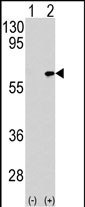
| Target/Specificity | This MAP3K7IP1 antibody is generated from rabbits immunized with a KLH conjugated synthetic peptide between 401-430 amino acids from human MAP3K7IP1. |
| Dilution | WB~~1:1000 E~~Use at an assay dependent concentration. |
| Format | Purified polyclonal antibody supplied in PBS with 0.09% (W/V) sodium azide. This antibody is purified through a protein A column, followed by peptide affinity purification. |
| Storage | Maintain refrigerated at 2-8°C for up to 2 weeks. For long term storage store at -20°C in small aliquots to prevent freeze-thaw cycles. |
| Precautions | MAP3K7IP1-S423 Antibody is for research use only and not for use in diagnostic or therapeutic procedures. |
| Name | TAB1 |
|---|---|
| Synonyms | MAP3K7IP1 |
| Function | Key adapter protein that plays an essential role in JNK and NF-kappa-B activation and proinflammatory cytokines production in response to stimulation with TLRs and cytokines (PubMed:22307082, PubMed:24403530). Mechanistically, associates with the catalytic domain of MAP3K7/TAK1 to trigger MAP3K7/TAK1 autophosphorylation leading to its full activation (PubMed:10838074, PubMed:25260751, PubMed:37832545). Similarly, associates with MAPK14 and triggers its autophosphorylation and subsequent activation (PubMed:11847341, PubMed:29229647). In turn, MAPK14 phosphorylates TAB1 and inhibits MAP3K7/TAK1 activation in a feedback control mechanism (PubMed:14592977). Also plays a role in recruiting MAPK14 to the TAK1 complex for the phosphorylation of the TAB2 and TAB3 regulatory subunits (PubMed:18021073). |
| Cellular Location | Cytoplasm, cytosol. Endoplasmic reticulum membrane; Peripheral membrane protein; Cytoplasmic side. Note=Recruited to the endoplasmic reticulum following interaction with STING1 |
| Tissue Location | Ubiquitous.. |
For Research Use Only. Not For Use In Diagnostic Procedures.
Provided below are standard protocols that you may find useful for product applications.
BACKGROUND
MAP3K7IP1 was identified as a regulator of the MAP kinase kinase kinase MAP3K7/TAK1, which is known to mediate various intracellular signaling pathways, such as those induced by TGF beta, interleukin 1, and WNT-1. This protein interacts and thus activates TAK1 kinase. It has been shown that the C-terminal portion of this protein is sufficient for binding and activation of TAK1, while a portion of the N-terminus acts as a dominant-negative inhibitor of TGF beta, suggesting that this protein may function as a mediator between TGF beta receptors and TAK1. This protein can also interact with and activate the mitogen-activated protein kinase 14 (MAPK14/p38alpha), and thus represents an alternative activation pathway, in addition to the MAPKK pathways, which contributes to the biological responses of MAPK14 to various stimuli.
REFERENCES
Arch,R.H., et.al., Genes Dev. 12 (18), 2821-2830 (1998)
Yamaguchi,K., et.al., EMBO J. 18 (1), 179-187 (1999)
终于等到您。ABCEPTA(百远生物)抗体产品。
点击下方“我要评价 ”按钮提交您的反馈信息,您的反馈和评价是我们最宝贵的财富之一,
我们将在1-3个工作日内处理您的反馈信息。
如有疑问,联系:0512-88856768 tech-china@abcepta.com.


