


 癌症的基本特征包括细胞增殖、血管生成、迁移、凋亡逃避机制和细胞永生等。找到癌症发生过程中这些通路的关键标记物和对应的抗体用于检测至关重要。
 为您推荐一个泛素化位点预测神器——泛素化分析工具,可以为您的蛋白的泛素化位点作出预测和评分。
 细胞自噬受体图形绘图工具为你的蛋白的细胞受体结合位点作出预测和评分,识别结合到自噬通路中的蛋白是非常重要的,便于让我们理解自噬在正常生理、病理过程中的作用,如发育、细胞分化、神经退化性疾病、压力条件下、感染和癌症。
Renin Receptor Polyclonal Antibody
- 产品详情
- 实验流程
- 背景知识
Application
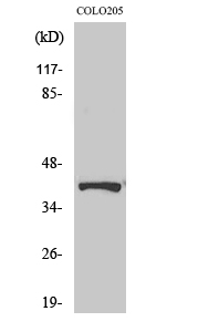
| WB, IHC-P, IF, ICC, E |
|---|---|
| Primary Accession | O75787 |
| Reactivity | Human, Mouse, Rat |
| Host | Rabbit |
| Clonality | Polyclonal |
| Calculated MW | 39008 Da |
| Gene ID | 10159 |
|---|---|
| Other Names | ATP6AP2; ATP6IP2; CAPER; ELDF10; HT028; MSTP009; PSEC0072; Renin receptor; ATPase H(+)-transporting lysosomal accessory protein 2; ATPase H(+)-transporting lysosomal-interacting protein 2; ER-localized type I transmembrane adaptor; Embryoni |
| Dilution | WB~~Western Blot: 1/500 - 1/2000. Immunohistochemistry: 1/100 - 1/300. Immunofluorescence: 1/200 - 1/1000. ELISA: 1/20000. Not yet tested in other applications. IHC-P~~1:50~200 IF~~1:50~200 ICC~~N/A E~~N/A |
| Format | Liquid in PBS containing 50% glycerol, 0.5% BSA and 0.09% (W/V) sodium azide. |
| Storage Conditions | -20℃ |
| Name | ATP6AP2 (HGNC:18305) |
|---|---|
| Function | Multifunctional protein which functions as a renin, prorenin cellular receptor and is involved in the assembly of the lysosomal proton-transporting V-type ATPase (V-ATPase) and the acidification of the endo-lysosomal system (PubMed:12045255, PubMed:29127204, PubMed:30374053, PubMed:32276428). May mediate renin-dependent cellular responses by activating ERK1 and ERK2 (PubMed:12045255). By increasing the catalytic efficiency of renin in AGT/angiotensinogen conversion to angiotensin I, may also play a role in the renin-angiotensin system (RAS) (PubMed:12045255). Through its function in V-type ATPase (v- ATPase) assembly and acidification of the lysosome it regulates protein degradation and may control different signaling pathways important for proper brain development, synapse morphology and synaptic transmission (By similarity). |
| Cellular Location | Endoplasmic reticulum membrane; Single-pass type I membrane protein. Lysosome membrane; Single- pass type I membrane protein. Cytoplasmic vesicle, autophagosome membrane {ECO:0000250|UniProtKB:Q9CYN9}; Single-pass type I membrane protein. Cell projection, dendritic spine membrane {ECO:0000250|UniProtKB:Q9CYN9}; Single-pass type I membrane protein. Cell projection, axon {ECO:0000250|UniProtKB:Q9CYN9}. Endosome membrane {ECO:0000250|UniProtKB:Q9CYN9}; Single-pass type I membrane protein. Cytoplasmic vesicle, clathrin-coated vesicle membrane {ECO:0000250|UniProtKB:Q6AXS4}; Single-pass type I membrane protein. Cytoplasmic vesicle, secretory vesicle, synaptic vesicle membrane {ECO:0000250|UniProtKB:Q6AXS4}; Single-pass type I membrane protein |
| Tissue Location | Expressed in brain, heart, placenta, liver, kidney and pancreas. Barely detectable in lung and skeletal muscles. In the kidney cortex it is restricted to the mesangium of glomeruli. In the coronary and kidney artery it is expressed in the subendothelium, associated to smooth muscles where it colocalizes with REN. Expressed in vascular structures and by syncytiotrophoblast cells in the mature fetal placenta. |
Research Areas
For Research Use Only. Not For Use In Diagnostic Procedures.
Application Protocols
Provided below are standard protocols that you may find useful for product applications.
BACKGROUND
Functions as a renin and prorenin cellular receptor. May mediate renin-dependent cellular responses by activating ERK1 and ERK2. By increasing the catalytic efficiency of renin in AGT/angiotensinogen conversion to angiotensin I, it may also play a role in the renin-angiotensin system (RAS).
终于等到您。ABCEPTA(百远生物)抗体产品。
点击下方“我要评价 ”按钮提交您的反馈信息,您的反馈和评价是我们最宝贵的财富之一,
我们将在1-3个工作日内处理您的反馈信息。
如有疑问,联系:0512-88856768 tech-china@abcepta.com.
¥ 1,500.00
Cat# AP72231


