


 癌症的基本特征包括细胞增殖、血管生成、迁移、凋亡逃避机制和细胞永生等。找到癌症发生过程中这些通路的关键标记物和对应的抗体用于检测至关重要。
 为您推荐一个泛素化位点预测神器——泛素化分析工具,可以为您的蛋白的泛素化位点作出预测和评分。
 细胞自噬受体图形绘图工具为你的蛋白的细胞受体结合位点作出预测和评分,识别结合到自噬通路中的蛋白是非常重要的,便于让我们理解自噬在正常生理、病理过程中的作用,如发育、细胞分化、神经退化性疾病、压力条件下、感染和癌症。
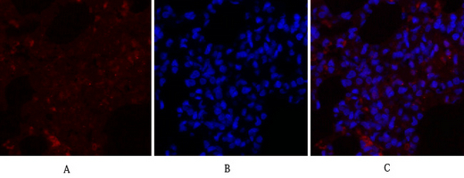
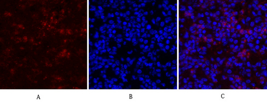
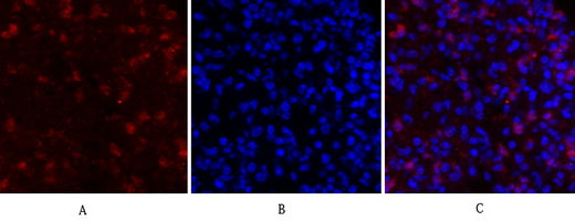
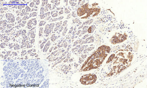
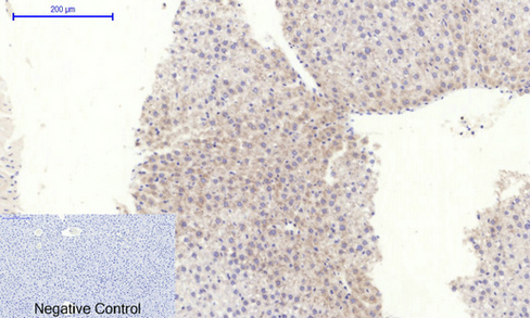
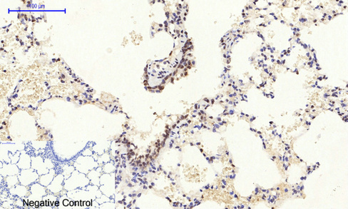
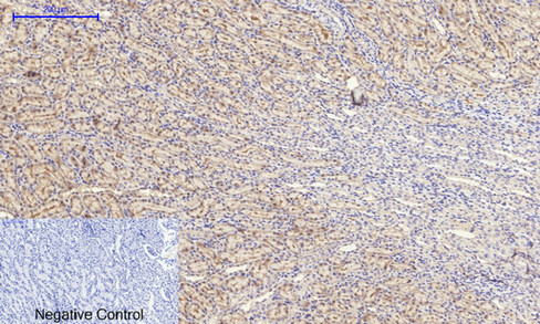
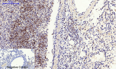
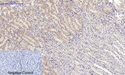
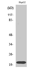
Rho A Polyclonal Antibody

- 产品详情
- 实验流程
- 背景知识
Application
| WB, IHC-P, IF |
|---|---|
| Primary Accession | P61586 |
| Reactivity | Human, Mouse, Rat |
| Host | Rabbit |
| Clonality | Polyclonal |
| Calculated MW | 21768 Da |
| Gene ID | 387 |
|---|---|
| Other Names | RHOA; ARH12; ARHA; RHO12; Transforming protein RhoA; Rho cDNA clone 12; h12 |
| Dilution | WB~~IF: 1:50-200 Western Blot: 1/500 - 1/2000. Immunohistochemistry: 1/100 - 1/300. ELISA: 1/5000. Not yet tested in other applications. IHC-P~~IF: 1:50-200 Western Blot: 1/500 - 1/2000. Immunohistochemistry: 1/100 - 1/300. ELISA: 1/5000. Not yet tested in other applications. IF~~IF: 1:50-200 Western Blot: 1/500 - 1/2000. Immunohistochemistry: 1/100 - 1/300. ELISA: 1/5000. Not yet tested in other applications. |
| Format | Liquid in PBS containing 50% glycerol, 0.5% BSA and 0.09% (W/V) sodium azide. |
| Storage Conditions | -20℃ |
| Name | RHOA (HGNC:667) |
|---|---|
| Synonyms | ARH12, ARHA, RHO12 |
| Function | Small GTPase which cycles between an active GTP-bound and an inactive GDP-bound state. Mainly associated with cytoskeleton organization, in active state binds to a variety of effector proteins to regulate cellular responses such as cytoskeletal dynamics, cell migration and cell cycle (PubMed:23871831). Regulates a signal transduction pathway linking plasma membrane receptors to the assembly of focal adhesions and actin stress fibers (PubMed:31570889, PubMed:8910519, PubMed:9121475). Involved in a microtubule-dependent signal that is required for the myosin contractile ring formation during cell cycle cytokinesis (PubMed:12900402, PubMed:16236794). Plays an essential role in cleavage furrow formation. Required for the apical junction formation of keratinocyte cell-cell adhesion (PubMed:20974804, PubMed:23940119). Essential for the SPATA13-mediated regulation of cell migration and adhesion assembly and disassembly (PubMed:19934221). The MEMO1-RHOA-DIAPH1 signaling pathway plays an important role in ERBB2- dependent stabilization of microtubules at the cell cortex. It controls the localization of APC and CLASP2 to the cell membrane, via the regulation of GSK3B activity. In turn, membrane-bound APC allows the localization of the MACF1 to the cell membrane, which is required for microtubule capture and stabilization (PubMed:20937854). Regulates KCNA2 potassium channel activity by reducing its location at the cell surface in response to CHRM1 activation; promotes KCNA2 endocytosis (PubMed:19403695, PubMed:9635436). Acts as an allosteric activator of guanine nucleotide exchange factor ECT2 by binding in its activated GTP-bound form to the PH domain of ECT2 which stimulates the release of PH inhibition and promotes the binding of substrate RHOA to the ECT2 catalytic center (PubMed:31888991). May be an activator of PLCE1 (PubMed:16103226). In neurons, involved in the inhibition of the initial spine growth. Upon activation by CaMKII, modulates dendritic spine structural plasticity by relaying CaMKII transient activation to synapse-specific, long-term signaling (By similarity). Acts as a regulator of platelet alpha-granule release during activation and aggregation of platelets (By similarity). When activated by DAAM1 may signal centrosome maturation and chromosomal segregation during cell division. May also be involved in contractile ring formation during cytokinesis. |
| Cellular Location | Cell membrane; Lipid-anchor; Cytoplasmic side. Cytoplasm, cytoskeleton. Cleavage furrow. Cytoplasm, cell cortex. Midbody. Cell projection, lamellipodium {ECO:0000250|UniProtKB:Q9QUI0}. Cell projection, dendrite {ECO:0000250|UniProtKB:Q9QUI0}. Nucleus Cytoplasm. Note=Localized to cell-cell contacts in calcium-treated keratinocytes (By similarity). Translocates to the equatorial region before furrow formation in a ECT2-dependent manner. Localizes to the equatorial cell cortex (at the site of the presumptive furrow) in early anaphase in an activated form and in a myosin- and actin-independent manner. Colocalizes with KANK1 at the contractile ring. Colocalizes with DAAM1 and KANK1 around centrosomes {ECO:0000250|UniProtKB:Q9QUI0} |
For Research Use Only. Not For Use In Diagnostic Procedures.
Provided below are standard protocols that you may find useful for product applications.
BACKGROUND
Regulates a signal transduction pathway linking plasma membrane receptors to the assembly of focal adhesions and actin stress fibers. Involved in a microtubule-dependent signal that is required for the myosin contractile ring formation during cell cycle cytokinesis. Plays an essential role in cleavage furrow formation. Required for the apical junction formation of keratinocyte cell-cell adhesion. Stimulates PKN2 kinase activity. May be an activator of PLCE1. Activated by ARHGEF2, which promotes the exchange of GDP for GTP. Essential for the SPATA13-mediated regulation of cell migration and adhesion assembly and disassembly. The MEMO1-RHOA-DIAPH1 signaling pathway plays an important role in ERBB2-dependent stabilization of microtubules at the cell cortex. It controls the localization of APC and CLASP2 to the cell membrane, via the regulation of GSK3B activity. In turn, membrane-bound APC allows the localization of the MACF1 to the cell membrane, which is required for microtubule capture and stabilization. Regulates a signal transduction pathway linking plasma membrane receptors to the assembly of focal adhesions and actin stress fibers. Involved in a microtubule-dependent signal that is required for the myosin contractile ring formation during cell cycle cytokinesis. Plays an essential role in cleavage furrow formation. Required for the apical junction formation of keratinocyte cell-cell adhesion. May be an activator of PLCE1. Activated by ARHGEF2, which promotes the exchange of GDP for GTP. Essential for the SPATA13-mediated regulation of cell migration and adhesion assembly and disassembly. The MEMO1-RHOA-DIAPH1 signaling pathway plays an important role in ERBB2-dependent stabilization of microtubules at the cell cortex. It controls the localization of APC and CLASP2 to the cell membrane, via the regulation of GSK3B activity. In turn, membrane-bound APC allows the localization of the MACF1 to the cell membrane, which is required for microtubule capture and stabilization (By similarity). Regulates KCNA2 potassium channel activity by reducing its location at the cell surface in response to CHRM1 activation; promotes KCNA2 endocytosis (PubMed:9635436, PubMed:19403695).
终于等到您。ABCEPTA(百远生物)抗体产品。
点击下方“我要评价 ”按钮提交您的反馈信息,您的反馈和评价是我们最宝贵的财富之一,
我们将在1-3个工作日内处理您的反馈信息。
如有疑问,联系:0512-88856768 tech-china@abcepta.com.


