


 癌症的基本特征包括细胞增殖、血管生成、迁移、凋亡逃避机制和细胞永生等。找到癌症发生过程中这些通路的关键标记物和对应的抗体用于检测至关重要。
 为您推荐一个泛素化位点预测神器——泛素化分析工具,可以为您的蛋白的泛素化位点作出预测和评分。
 细胞自噬受体图形绘图工具为你的蛋白的细胞受体结合位点作出预测和评分,识别结合到自噬通路中的蛋白是非常重要的,便于让我们理解自噬在正常生理、病理过程中的作用,如发育、细胞分化、神经退化性疾病、压力条件下、感染和癌症。
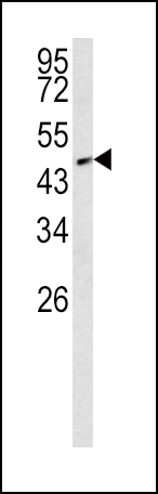
SMAD2 Antibody (T220)
Affinity Purified Rabbit Polyclonal Antibody (Pab)
_-_SY5Y.jpg)
- 产品详情
- 实验流程
- 背景知识
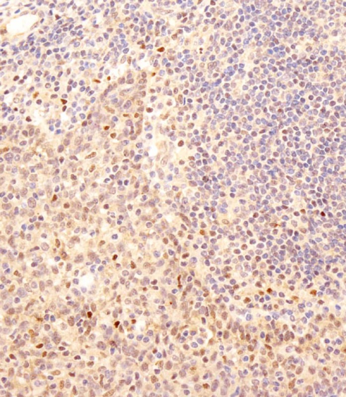
Application
| WB, IF, IHC-P-Leica, E |
|---|---|
| Primary Accession | Q15796 |
| Other Accession | O70436, Q62432, Q9I9P9, Q1W668 |
| Reactivity | Human, Mouse, Rat |
| Predicted | Mouse, Rat, Zebrafish, Bovine |
| Host | Rabbit |
| Clonality | Polyclonal |
| Isotype | Rabbit IgG |
| Calculated MW | 52306 Da |
| Antigen Region | 201-230 aa |
| Gene ID | 4087 |
|---|---|
| Other Names | Mothers against decapentaplegic homolog 2, MAD homolog 2, Mothers against DPP homolog 2, JV18-1, Mad-related protein 2, hMAD-2, SMAD family member 2, SMAD 2, Smad2, hSMAD2, SMAD2, MADH2, MADR2 |
| Target/Specificity | This SMAD2 antibody is generated from rabbits immunized with a KLH conjugated synthetic peptide between 201-230 amino acids from human SMAD2. |
| Dilution | WB~~1:1000 IF~~1:200 IHC-P-Leica~~1:250 E~~Use at an assay dependent concentration. |
| Format | Purified polyclonal antibody supplied in PBS with 0.09% (W/V) sodium azide. This antibody is purified through a protein A column, followed by peptide affinity purification. |
| Storage | Maintain refrigerated at 2-8°C for up to 2 weeks. For long term storage store at -20°C in small aliquots to prevent freeze-thaw cycles. |
| Precautions | SMAD2 Antibody (T220) is for research use only and not for use in diagnostic or therapeutic procedures. |
| Name | SMAD2 (HGNC:6768) |
|---|---|
| Synonyms | MADH2, MADR2 |
| Function | Receptor-regulated SMAD (R-SMAD) that is an intracellular signal transducer and transcriptional modulator activated by TGF-beta (transforming growth factor) and activin type 1 receptor kinases. Binds the TRE element in the promoter region of many genes that are regulated by TGF-beta and, on formation of the SMAD2/SMAD4 complex, activates transcription. Promotes TGFB1-mediated transcription of odontoblastic differentiation genes in dental papilla cells (By similarity). Positively regulates PDPK1 kinase activity by stimulating its dissociation from the 14-3-3 protein YWHAQ which acts as a negative regulator. May act as a tumor suppressor in colorectal carcinoma (PubMed:8752209). |
| Cellular Location | Cytoplasm. Nucleus. Note=Cytoplasmic and nuclear in the absence of TGF-beta. On TGF-beta stimulation, migrates to the nucleus when complexed with SMAD4 or with IPO7 (PubMed:21145499, PubMed:9865696). On dephosphorylation by phosphatase PPM1A, released from the SMAD2/SMAD4 complex, and exported out of the nucleus by interaction with RANBP1 (PubMed:16751101, PubMed:19289081). Localized mainly to the nucleus in the early stages of embryo development with expression becoming evident in the cytoplasm at the blastocyst and epiblast stages (By similarity). {ECO:0000250|UniProtKB:Q62432, ECO:0000269|PubMed:16751101, ECO:0000269|PubMed:19289081, ECO:0000269|PubMed:21145499, ECO:0000269|PubMed:9865696} |
| Tissue Location | Expressed at high levels in skeletal muscle, endothelial cells, heart and placenta. |
For Research Use Only. Not For Use In Diagnostic Procedures.
Provided below are standard protocols that you may find useful for product applications.
BACKGROUND
The protein belongs to the SMAD, a family of proteins similar to the proteins of the Drosophila gene 'mothers against decapentaplegic' (Mad) and the C. elegans gene Sma. SMAD proteins are signal transducers and transcriptional modulators that mediate multiple signaling pathways. This protein mediates the signal of the transforming growth factor (TGF)-beta, and thus regulates multiple cellular processes, such as cell proliferation, apoptosis, and differentiation. This protein is recruited to the TGF-beta receptors through its interaction with the SMAD anchor for receptor activation (SARA) protein. In response to TGF-beta signal, this protein is phosphorylated by the TGF-beta receptors. The phosphorylation induces the dissociation of this protein with SARA and the association with the family member SMAD4. The association with SMAD4 is important for the translocation of this protein into the nucleus, where it binds to target promoters and forms a transcription repressor complex with other cofactors. This protein can also be phosphorylated by activin type 1 receptor kinase, and mediates the signal from the activin.
REFERENCES
References for protein:
1.Funaba,M., J. Biol. Chem. 277 (44), 41361-41368 (2002)
2.Wicks,S.J., Mol. Cell. Biol. 20 (21), 8103-8111 (2000)
References for SY5Y (SH-SY5Y; ATCC#CRL-2266): 1. Ross RA, et al. Coordinate morphological and biochemical interconversion of human neuroblastoma cells. J. Natl. Cancer Inst. 71: 741-749, 1983. [PubMed: 6137586]; 2. Biedler JL, et al. Multiple neurotransmitter synthesis by human neuroblastoma cell lines and clones. Cancer Res. 38: 3751-3757, 1978. [PubMed: 29704].
终于等到您。ABCEPTA(百远生物)抗体产品。
点击下方“我要评价 ”按钮提交您的反馈信息,您的反馈和评价是我们最宝贵的财富之一,
我们将在1-3个工作日内处理您的反馈信息。
如有疑问,联系:0512-88856768 tech-china@abcepta.com.


