


 癌症的基本特征包括细胞增殖、血管生成、迁移、凋亡逃避机制和细胞永生等。找到癌症发生过程中这些通路的关键标记物和对应的抗体用于检测至关重要。
 为您推荐一个泛素化位点预测神器——泛素化分析工具,可以为您的蛋白的泛素化位点作出预测和评分。
 细胞自噬受体图形绘图工具为你的蛋白的细胞受体结合位点作出预测和评分,识别结合到自噬通路中的蛋白是非常重要的,便于让我们理解自噬在正常生理、病理过程中的作用,如发育、细胞分化、神经退化性疾病、压力条件下、感染和癌症。
DDB1 Rabbit mAb
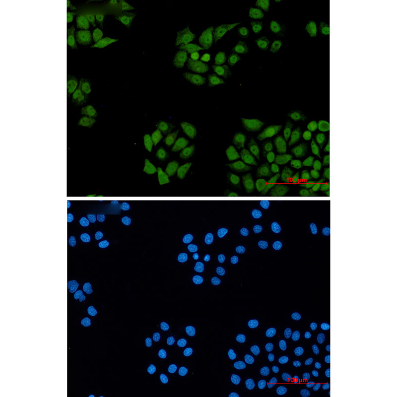

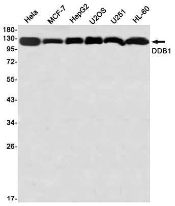
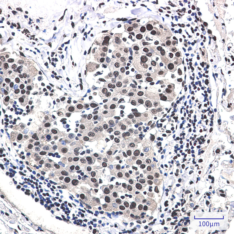
- 产品详情
- 实验流程
- 背景知识
Application 
| WB, IHC-P |
|---|---|
| Primary Accession | Q16531 |
| Reactivity | Rat, Human, Mouse |
| Host | Rabbit |
| Clonality | Monoclonal Antibody |
| Isotype | IgG |
| Conjugate | Unconjugated |
| Purification | Affinity Purified |
| Calculated MW | 126968 Da |
| Gene ID | 1642 |
|---|---|
| Other Names | DDB1 |
| Dilution | WB~~1:1000-1:5000 IHC-P~~1:50~200 |
| Format | Liquid in 50mM Tris-Glycine(pH 7.4), 0.15M NaCl, 40%Glycerol, 0.01% sodium azide and 0.05% BSA. |
| Storage | Store at 4°C short term. Aliquot and store at -20°C long term. Avoid freeze/thaw cycles. |
For Research Use Only. Not For Use In Diagnostic Procedures.
| Name | DDB1 |
|---|---|
| Synonyms | XAP1 |
| Function | Involved in DNA repair and protein ubiquitination, as part of the UV-DDB complex and DCX (DDB1-CUL4-X-box) complexes, respectively (PubMed:14739464, PubMed:15448697, PubMed:16260596, PubMed:16407242, PubMed:16407252, PubMed:16482215, PubMed:16940174, PubMed:17079684, PubMed:25970626). Core component of the UV-DDB complex (UV-damaged DNA- binding protein complex), a complex that recognizes UV-induced DNA damage and recruits proteins of the nucleotide excision repair pathway (the NER pathway) to initiate DNA repair (PubMed:15448697, PubMed:16260596, PubMed:16407242, PubMed:16940174). The UV-DDB complex preferentially binds to cyclobutane pyrimidine dimers (CPD), 6-4 photoproducts (6-4 PP), apurinic sites and short mismatches (PubMed:15448697, PubMed:16260596, PubMed:16407242, PubMed:16940174). Component of numerous distinct DCX (DDB1-CUL4-X-box) E3 ubiquitin- protein ligase complexes which mediate the ubiquitination and subsequent proteasomal degradation of target proteins (PubMed:14739464, PubMed:16407252, PubMed:16482215, PubMed:17079684, PubMed:18332868, PubMed:18381890, PubMed:19966799, PubMed:22118460, PubMed:25043012, PubMed:25108355, PubMed:28886238, PubMed:41565821). The functional specificity of the DCX E3 ubiquitin-protein ligase complex is determined by the variable substrate recognition component recruited by DDB1 (PubMed:14739464, PubMed:16407252, PubMed:16482215, PubMed:17079684, PubMed:18332868, PubMed:18381890, PubMed:19966799, PubMed:22118460, PubMed:25043012, PubMed:25108355). DCX(DDB2) (also known as DDB1-CUL4-ROC1, CUL4-DDB-ROC1 and CUL4-DDB-RBX1) may ubiquitinate histone H2A, histone H3 and histone H4 at sites of UV- induced DNA damage (PubMed:16473935, PubMed:16678110, PubMed:17041588, PubMed:18593899). The ubiquitination of histones may facilitate their removal from the nucleosome and promote subsequent DNA repair (PubMed:16473935, PubMed:16678110, PubMed:17041588, PubMed:18593899). DCX(DDB2) also ubiquitinates XPC, which may enhance DNA-binding by XPC and promote NER (PubMed:15882621). DCX(DTL) plays a role in PCNA- dependent polyubiquitination of CDT1 and MDM2-dependent ubiquitination of TP53 in response to radiation-induced DNA damage and during DNA replication (PubMed:17041588). DCX(ERCC8) (the CSA complex) plays a role in transcription-coupled repair (TCR) (PubMed:12732143, PubMed:32355176, PubMed:38316879). The DDB1-CUL4A-DTL E3 ligase complex regulates the circadian clock function by mediating the ubiquitination and degradation of CRY1 (PubMed:26431207). DDB1-mediated CRY1 degradation promotes FOXO1 protein stability and FOXO1-mediated gluconeogenesis in the liver (By similarity). By acting on TET dioxygenases, essential for oocyte maintenance at the primordial follicle stage, hence essential for female fertility (By similarity). Maternal factor required for proper zygotic genome activation and genome reprogramming (By similarity). |
| Cellular Location | Cytoplasm. Nucleus. Note=Primarily cytoplasmic (PubMed:10777491, PubMed:11673459). Translocates to the nucleus following UV irradiation and subsequently accumulates at sites of DNA damage (PubMed:10777491, PubMed:11673459). More concentrated in nuclei than in cytoplasm in germinal vesicle (GV) stage oocytes, zygotes and the 2-cell stage, but distributed in the cytoplasm at the MII-stage oocytes (By similarity). {ECO:0000250|UniProtKB:Q3U1J4, ECO:0000269|PubMed:10777491, ECO:0000269|PubMed:11673459} |
Provided below are standard protocols that you may find useful for product applications.
BACKGROUND
The protein encoded by this gene is the large subunit (p127) of the heterodimeric DNA damage-binding (DDB) complex while another protein (p48) forms the small subunit. This protein complex functions in nucleotide-excision repair and binds to DNA following UV damage. Defective activity of this complex causes the repair defect in patients with xeroderma pigmentosum complementation group E (XPE) - an autosomal recessive disorder characterized by photosensitivity and early onset of carcinomas. However, it remains for mutation analysis to demonstrate whether the defect in XPE patients is in this gene or the gene encoding the small subunit. In addition, Best vitelliform mascular dystrophy is mapped to the same region as this gene on 11q, but no sequence alternations of this gene are demonstrated in Best disease patients. The protein encoded by this gene also functions as an adaptor molecule for the cullin 4 (CUL4) ubiquitin E3 ligase complex by facilitating the binding of substrates to this complex and the ubiquitination of proteins.

终于等到您。ABCEPTA(百远生物)抗体产品。
点击下方“我要评价 ”按钮提交您的反馈信息,您的反馈和评价是我们最宝贵的财富之一,
我们将在1-3个工作日内处理您的反馈信息。
如有疑问,联系:0512-88856768 tech-china@abcepta.com.


