


 癌症的基本特征包括细胞增殖、血管生成、迁移、凋亡逃避机制和细胞永生等。找到癌症发生过程中这些通路的关键标记物和对应的抗体用于检测至关重要。
 为您推荐一个泛素化位点预测神器——泛素化分析工具,可以为您的蛋白的泛素化位点作出预测和评分。
 细胞自噬受体图形绘图工具为你的蛋白的细胞受体结合位点作出预测和评分,识别结合到自噬通路中的蛋白是非常重要的,便于让我们理解自噬在正常生理、病理过程中的作用,如发育、细胞分化、神经退化性疾病、压力条件下、感染和癌症。
MYH9 Rabbit mAb
- 产品详情
- 实验流程
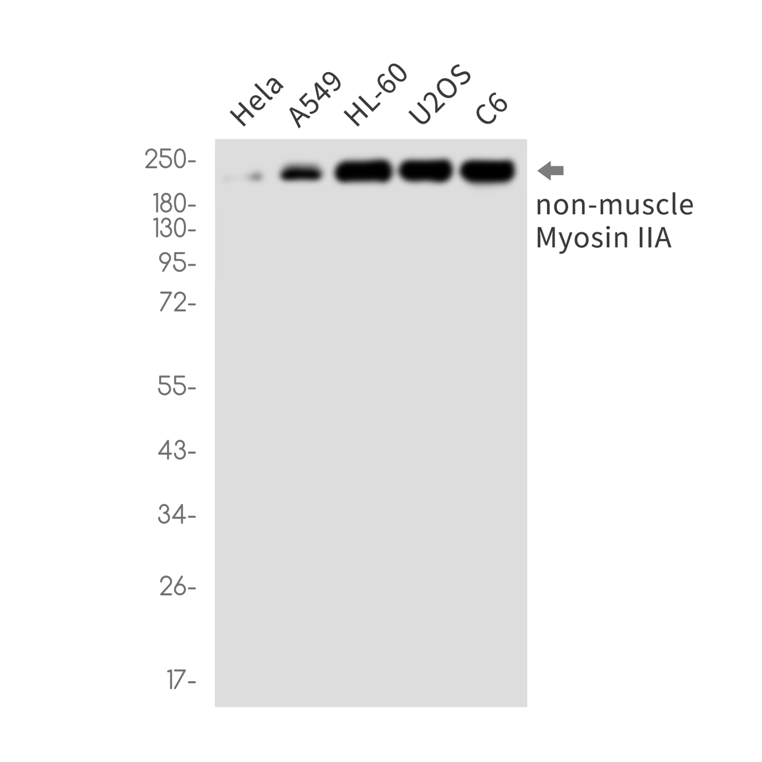
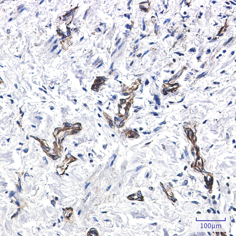
Application
| WB, IHC-P, IP |
|---|---|
| Primary Accession | P35579 |
| Reactivity | Human, Rat |
| Host | Rabbit |
| Clonality | Monoclonal Antibody |
| Calculated MW | 226532 Da |
| Gene ID | 4627 |
|---|---|
| Other Names | MYH9 |
| Dilution | WB~~1/500-1/1000 IHC-P~~1:50~200 IP~~N/A |
| Format | 50mM Tris-Glycine(pH 7.4), 0.15M NaCl, 40%Glycerol, 0.01% sodium azide and 0.05% BSA. |
| Storage | Store at 4°C short term. Aliquot and store at -20°C long term. Avoid freeze/thaw cycles. |
| Name | MYH9 |
|---|---|
| Function | Cellular myosin that appears to play a role in cytokinesis, cell shape, and specialized functions such as secretion and capping. Required for cortical actin clearance prior to oocyte exocytosis (By similarity). Promotes cell motility in conjunction with S100A4 (PubMed:16707441). During cell spreading, plays an important role in cytoskeleton reorganization, focal contact formation (in the margins but not the central part of spreading cells), and lamellipodial retraction; this function is mechanically antagonized by MYH10 (PubMed:20052411). |
| Cellular Location | Cytoplasm, cytoskeleton. Cytoplasm, cell cortex {ECO:0000250|UniProtKB:Q8VDD5}. Cytoplasmic vesicle, secretory vesicle, Cortical granule {ECO:0000250|UniProtKB:Q8VDD5}. Cell membrane Note=Colocalizes with actin filaments at lamellipodia margins and at the leading edge of migrating cells (PubMed:20052411). In retinal pigment epithelial cells, predominantly localized to stress fiber-like structures with some localization to cytoplasmic puncta (PubMed:27331610). |
| Tissue Location | In the kidney, expressed in the glomeruli. Also expressed in leukocytes. |
Research Areas
For Research Use Only. Not For Use In Diagnostic Procedures.
Application Protocols
Provided below are standard protocols that you may find useful for product applications.
终于等到您。ABCEPTA(百远生物)抗体产品。
点击下方“我要评价 ”按钮提交您的反馈信息,您的反馈和评价是我们最宝贵的财富之一,
我们将在1-3个工作日内处理您的反馈信息。
如有疑问,联系:0512-88856768 tech-china@abcepta.com.
¥ 1,500.00
Cat# AP76621


