


 癌症的基本特征包括细胞增殖、血管生成、迁移、凋亡逃避机制和细胞永生等。找到癌症发生过程中这些通路的关键标记物和对应的抗体用于检测至关重要。
 为您推荐一个泛素化位点预测神器——泛素化分析工具,可以为您的蛋白的泛素化位点作出预测和评分。
 细胞自噬受体图形绘图工具为你的蛋白的细胞受体结合位点作出预测和评分,识别结合到自噬通路中的蛋白是非常重要的,便于让我们理解自噬在正常生理、病理过程中的作用,如发育、细胞分化、神经退化性疾病、压力条件下、感染和癌症。
DTX1 Antibody (Center) 精选
Affinity Purified Rabbit Polyclonal Antibody (Pab)
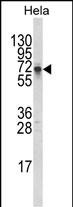
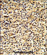
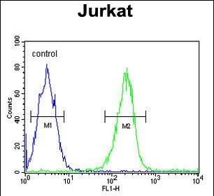
- 产品详情
- 实验流程
- 背景知识
Application 
| WB, IHC-P, FC, E |
|---|---|
| Primary Accession | Q86Y01 |
| Other Accession | Q61010 |
| Reactivity | Human |
| Predicted | Mouse, Rat, Bovine, Canine, Chicken |
| Host | Rabbit |
| Clonality | Polyclonal |
| Isotype | Rabbit IgG |
| Calculated MW | 67368 Da |
| Antigen Region | 382-410 aa |
| Gene ID | 1840 |
|---|---|
| Other Names | E3 ubiquitin-protein ligase DTX1, 632-, Protein deltex-1, Deltex1, hDTX1, DTX1 |
| Target/Specificity | This DTX1 antibody is generated from rabbits immunized with a KLH conjugated synthetic peptide between 382-410 amino acids from the Central region of human DTX1. |
| Dilution | WB~~1:1000 IHC-P~~1:100~500 FC~~1:10~50 E~~Use at an assay dependent concentration. |
| Format | Purified polyclonal antibody supplied in PBS with 0.09% (W/V) sodium azide. This antibody is purified through a protein A column, followed by peptide affinity purification. |
| Storage | Maintain refrigerated at 2-8°C for up to 2 weeks. For long term storage store at -20°C in small aliquots to prevent freeze-thaw cycles. |
| Precautions | DTX1 Antibody (Center) is for research use only and not for use in diagnostic or therapeutic procedures. |
For Research Use Only. Not For Use In Diagnostic Procedures.
| Name | DTX1 |
|---|---|
| Function | E3 ubiquitin-protein ligase that mediates ubiquitination and proteasomal degradation of MEKK1, thereby modulating signaling pathways involved in cell fate determination (By similarity). Regulates the Notch signaling pathway, acting predominantly as a positive regulator but functioning as a context-dependent negative regulator in specific developmental or cellular settings. Mediates the antineural activity of Notch signaling, likely by inhibiting transcriptional activation mediated by MATCH1. Negatively regulates Notch signaling by controlling the activity of PIP4K2C, a lipid kinase that promotes recycling of Notch receptors from RAB4A-positive endosomes to the cell surface (PubMed:29440432). Participates in several developmental and immune processes, including neurogenesis, lymphogenesis, and myogenesis. Influences lymphocyte differentiation by promoting B-cell development at the expense of T-cell development, suggesting functional antagonism of NOTCH1 in this context. Also promotes degradation of PRKCQ via the endosome-lysosome pathway, contributing to PKCtheta down-regulation during T-cell anergy (PubMed:25000980). In addition, facilitates differentiation of Th17 cells by inducing expression and DNA-binding activity of RORgT (PubMed:32736174). |
| Cellular Location | Cytoplasm. Nucleus. Endosome Note=Predominantly cytoplasmic. Associates with endocytic vesicles Partially nuclear. |
| Tissue Location | Widely expressed. Strongly expressed in blood vessel. Also expressed in embryonic nervous system, pancreas, lung, adrenal gland, digestive tube and muscles. Expressed in MZB cells and developing B- and T-cells. |
Research Areas
Application Protocols
Provided below are standard protocols that you may find useful for product applications.
BACKGROUND
DTX1 was identified as encoding a positive regulator of the Notch-signaling pathway in Drosophila. The human gene encodes a protein of unknown function; however, it may play a role in basic helix-loop-helix transcription factor activity.
REFERENCES
Wu,C.,et.al., Proteomics 7 (11), 1775-1785 (2007)

终于等到您。ABCEPTA(百远生物)抗体产品。
点击下方“我要评价 ”按钮提交您的反馈信息,您的反馈和评价是我们最宝贵的财富之一,
我们将在1-3个工作日内处理您的反馈信息。
如有疑问,联系:0512-88856768 tech-china@abcepta.com.
¥ 1,250.00
Cat# AP8923c


