


 癌症的基本特征包括细胞增殖、血管生成、迁移、凋亡逃避机制和细胞永生等。找到癌症发生过程中这些通路的关键标记物和对应的抗体用于检测至关重要。
 为您推荐一个泛素化位点预测神器——泛素化分析工具,可以为您的蛋白的泛素化位点作出预测和评分。
 细胞自噬受体图形绘图工具为你的蛋白的细胞受体结合位点作出预测和评分,识别结合到自噬通路中的蛋白是非常重要的,便于让我们理解自噬在正常生理、病理过程中的作用,如发育、细胞分化、神经退化性疾病、压力条件下、感染和癌症。
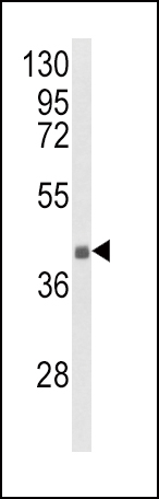
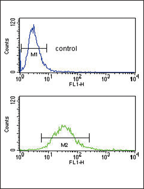
HLA-G Antibody (Center)
Affinity Purified Rabbit Polyclonal Antibody (Pab)

- 产品详情
- 文献引用 : 5
- 实验流程
- 背景知识
Application
| IHC-P-Leica, FC, WB, E |
|---|---|
| Primary Accession | P17693 |
| Reactivity | Human |
| Host | Rabbit |
| Clonality | Polyclonal |
| Isotype | Rabbit IgG |
| Calculated MW | 38224 Da |
| Antigen Region | 62-89 aa |
| Gene ID | 3135 |
|---|---|
| Other Names | HLA class I histocompatibility antigen, alpha chain G, HLA G antigen, MHC class I antigen G, HLA-G, HLA-60, HLAG |
| Target/Specificity | This HLA-G antibody is generated from rabbits immunized with a KLH conjugated synthetic peptide between 62-89 amino acids from the Central region of human HLA-G. |
| Dilution | IHC-P-Leica~~1:500 FC~~1:10~50 WB~~1:1000 E~~Use at an assay dependent concentration. |
| Format | Purified polyclonal antibody supplied in PBS with 0.09% (W/V) sodium azide. This antibody is purified through a protein A column, followed by peptide affinity purification. |
| Storage | Maintain refrigerated at 2-8°C for up to 2 weeks. For long term storage store at -20°C in small aliquots to prevent freeze-thaw cycles. |
| Precautions | HLA-G Antibody (Center) is for research use only and not for use in diagnostic or therapeutic procedures. |
| Name | HLA-G {ECO:0000303|PubMed:1570318, ECO:0000312|HGNC:HGNC:4964} |
|---|---|
| Function | [Isoform 1]: Non-classical major histocompatibility class Ib molecule involved in immune regulatory processes at the maternal-fetal interface (PubMed:19304799, PubMed:23184984, PubMed:29262349). In complex with B2M/beta-2 microglobulin binds a limited repertoire of nonamer self-peptides derived from intracellular proteins including histones and ribosomal proteins (PubMed:7584149, PubMed:8805247). Peptide-bound HLA-G-B2M complex acts as a ligand for inhibitory/activating KIR2DL4, LILRB1 and LILRB2 receptors on uterine immune cells to promote fetal development while maintaining maternal- fetal tolerance (PubMed:16366734, PubMed:19304799, PubMed:20448110, PubMed:23184984, PubMed:27859042, PubMed:29262349). Upon interaction with KIR2DL4 and LILRB1 receptors on decidual NK cells, it triggers NK cell senescence-associated secretory phenotype as a molecular switch to promote vascular remodeling and fetal growth in early pregnancy (PubMed:16366734, PubMed:19304799, PubMed:23184984, PubMed:29262349). Through interaction with KIR2DL4 receptor on decidual macrophages induces pro-inflammatory cytokine production mainly associated with tissue remodeling (PubMed:19304799). Through interaction with LILRB2 receptor triggers differentiation of type 1 regulatory T cells and myeloid-derived suppressor cells, both of which actively maintain maternal-fetal tolerance (PubMed:20448110, PubMed:27859042). May play a role in balancing tolerance and antiviral-immunity at maternal-fetal interface by keeping in check the effector functions of NK, CD8+ T cells and B cells (PubMed:10190900, PubMed:11290782, PubMed:24453251). Reprograms B cells toward an immune suppressive phenotype via LILRB1 (PubMed:24453251). May induce immune activation/suppression via intercellular membrane transfer (trogocytosis), likely enabling interaction with KIR2DL4, which resides mostly in endosomes (PubMed:20179272, PubMed:26460007). Through interaction with the inhibitory receptor CD160 on endothelial cells may control angiogenesis in immune privileged sites (PubMed:16809620). |
| Cellular Location | [Isoform 1]: Cell membrane; Single-pass type I membrane protein. Endoplasmic reticulum membrane. Early endosome membrane [Isoform 2]: Cell membrane; Single-pass type I membrane protein [Isoform 4]: Cell membrane; Single-pass type I membrane protein [Isoform 6]: Secreted Cell projection, filopodium membrane. Note=HLA-G trogocytosis from extravillous trophoblast's filopodia occurs in the majority of decidual NK cells. |
| Tissue Location | Expressed in adult eye (PubMed:1570318). Expressed in immune cell subsets including monocytes, myeloid and plasmacytoid dendritic cells and regulatory T cells (Tr1)(at protein level) (PubMed:20448110). Secreted by follicular dendritic cell and follicular helper T cells (PubMed:24453251) [Isoform 7]: Expressed in placenta, amniotic membrane, skin, cord blood and peripheral blood mononuclear cells |
For Research Use Only. Not For Use In Diagnostic Procedures.

Provided below are standard protocols that you may find useful for product applications.
BACKGROUND
HLA-G belongs to the HLA class I heavy chain paralogues. This class I molecule is a heterodimer consisting of a heavy chain and a light chain (beta-2 microglobulin). The heavy chain is anchored in the membrane. HLA-G is expressed on fetal derived placental cells. The heavy chain is approximately 45 kDa and its gene contains 8 exons. Exon one encodes the leader peptide, exons 2 and 3 encode the alpha1 and alpha2 domain, which both bind the peptide, exon 4 encodes the alpha3 domain, exon 5 encodes the transmembrane region, and exon 6 encodes the cytoplasmic tail.
REFERENCES
Walpole,N.G., et.al., J. Mol. Biol. 397 (2), 467-480 (2010)
Tian,W., et.sl., Tissue Antigens 75 (3), 227-234 (2010)
终于等到您。ABCEPTA(百远生物)抗体产品。
点击下方“我要评价 ”按钮提交您的反馈信息,您的反馈和评价是我们最宝贵的财富之一,
我们将在1-3个工作日内处理您的反馈信息。
如有疑问,联系:0512-88856768 tech-china@abcepta.com.


