


 癌症的基本特征包括细胞增殖、血管生成、迁移、凋亡逃避机制和细胞永生等。找到癌症发生过程中这些通路的关键标记物和对应的抗体用于检测至关重要。
 为您推荐一个泛素化位点预测神器——泛素化分析工具,可以为您的蛋白的泛素化位点作出预测和评分。
 细胞自噬受体图形绘图工具为你的蛋白的细胞受体结合位点作出预测和评分,识别结合到自噬通路中的蛋白是非常重要的,便于让我们理解自噬在正常生理、病理过程中的作用,如发育、细胞分化、神经退化性疾病、压力条件下、感染和癌症。
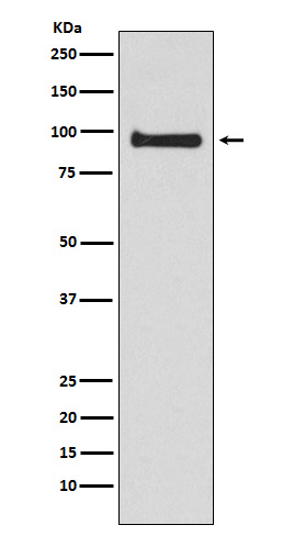
VAV3 Antibody
Rabbit mAb
- 产品详情
- 实验流程
Application
| WB, IF, FC, ICC, IP |
|---|---|
| Primary Accession | Q9UKW4 |
| Reactivity | Human, Mouse |
| Clonality | Monoclonal |
| Other Names | RGD1565941; VAV 3; Vav3; VAV3 oncogene; |
| Isotype | Rabbit IgG |
| Host | Rabbit |
| Calculated MW | 97776 Da |
| Dilution | WB 1:500~1:2000 ICC/IF 1:50~1:200 IP 1:50 FC 1:40. |
|---|---|
| Purification | Affinity-chromatography |
| Immunogen | A synthesized peptide derived from human VAV3 |
| Description | Plays an important role in angiogenesis. Its recruitement by phosphorylated EPHA2 is critical for EFNA1-induced RAC1 GTPase activation and vascular endothelial cell migration and assembly. |
| Storage Condition and Buffer | Rabbit IgG in phosphate buffered saline , pH 7.4, 150mM NaCl, 0.02% sodium azide and 50% glycerol. Store at +4°C short term. Store at -20°C long term. Avoid freeze / thaw cycle. |
| Name | VAV3 |
|---|---|
| Function | Exchange factor for GTP-binding proteins RhoA, RhoG and, to a lesser extent, Rac1. Binds physically to the nucleotide-free states of those GTPases. Plays an important role in angiogenesis. Its recruitment by phosphorylated EPHA2 is critical for EFNA1-induced RAC1 GTPase activation and vascular endothelial cell migration and assembly (By similarity). May be important for integrin-mediated signaling, at least in some cell types. In osteoclasts, along with SYK tyrosine kinase, required for signaling through integrin alpha-v/beta-1 (ITAGV-ITGB1), a crucial event for osteoclast proper cytoskeleton organization and function. This signaling pathway involves RAC1, but not RHO, activation. Necessary for proper wound healing. In the course of wound healing, required for the phagocytotic cup formation preceding macrophage phagocytosis of apoptotic neutrophils. Responsible for integrin beta-2 (ITGB2)-mediated macrophage adhesion and, to a lesser extent, contributes to beta-3 (ITGB3)-mediated adhesion. Does not affect integrin beta-1 (ITGB1)-mediated adhesion (By similarity). |
| Tissue Location | Isoform 1 and isoform 3 are widely expressed; both are expressed at very low levels in skeletal muscle. In keratinocytes, isoform 1 is less abundant than isoform 3. Isoform 3 is detected at very low levels, if any, in adrenal gland, bone marrow, spleen, fetal brain and spinal cord; in these tissues, isoform 1 is readily detectable. |
Research Areas
For Research Use Only. Not For Use In Diagnostic Procedures.
Application Protocols
Provided below are standard protocols that you may find useful for product applications.
终于等到您。ABCEPTA(百远生物)抗体产品。
点击下方“我要评价 ”按钮提交您的反馈信息,您的反馈和评价是我们最宝贵的财富之一,
我们将在1-3个工作日内处理您的反馈信息。
如有疑问,联系:0512-88856768 tech-china@abcepta.com.
¥ 1,500.00
Cat# AP92334


