


 癌症的基本特征包括细胞增殖、血管生成、迁移、凋亡逃避机制和细胞永生等。找到癌症发生过程中这些通路的关键标记物和对应的抗体用于检测至关重要。
 为您推荐一个泛素化位点预测神器——泛素化分析工具,可以为您的蛋白的泛素化位点作出预测和评分。
 细胞自噬受体图形绘图工具为你的蛋白的细胞受体结合位点作出预测和评分,识别结合到自噬通路中的蛋白是非常重要的,便于让我们理解自噬在正常生理、病理过程中的作用,如发育、细胞分化、神经退化性疾病、压力条件下、感染和癌症。
Recombinant Anti-MLCK1 Rabbit mAb
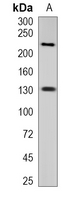
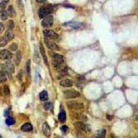
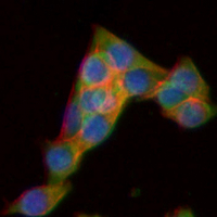
- 产品详情
- 实验流程
Application 
| WB, IHC, FC, IF/IC |
|---|---|
| Primary Accession | Q15746 |
| Reactivity | Human, Mouse, Rat |
| Host | Rabbit |
| Clonality | Monoclonal |
| Calculated MW | 210715 Da |
| Gene ID | 4638 |
|---|---|
| Other Names | MLCK; MLCK1; MYLK1; Myosin light chain kinase, smooth muscle; MLCK; smMLCK; Kinase-related protein; KRP; Telokin |
| Target/Specificity | KLH-conjugated synthetic peptide encompassing a sequence within human MLCK1 protein. The exact sequence is proprietary. |
| Dilution | WB~~WB (1/500 - 1/1000) IHC~~1:100~500 FC~~1:10~50 IF/IC~~N/A |
| Format | Liquid in PBS, pH 7.3, 50% glycerol, 0.05% BSA, and 0.05% Proclin300. |
| Storage | Store at -20 °C.Stable for 12 months from date of receipt |
For Research Use Only. Not For Use In Diagnostic Procedures.
| Name | MYLK (HGNC:7590) |
|---|---|
| Synonyms | MLCK, MLCK1, MYLK1 |
| Function | Calcium/calmodulin-dependent myosin light chain kinase implicated in smooth muscle contraction via phosphorylation of myosin light chains (MLC) (PubMed:11113114). Also regulates actin-myosin interaction through a non-kinase activity (PubMed:11113114). Phosphorylates PTK2B/PYK2 and myosin light-chains (PubMed:11113114, PubMed:18587400). Involved in the inflammatory response (e.g. apoptosis, vascular permeability, leukocyte diapedesis), cell motility and morphology, airway hyperreactivity and other activities relevant to asthma (PubMed:20139351). Required for tonic airway smooth muscle contraction that is necessary for physiological and asthmatic airway resistance (By similarity). Necessary for gastrointestinal motility (By similarity). Implicated in the regulation of endothelial as well as vascular permeability, probably via the regulation of cytoskeletal rearrangements (By similarity). In the nervous system it has been shown to control the growth initiation of astrocytic processes in culture and to participate in transmitter release at synapses formed between cultured sympathetic ganglion cells (PubMed:20375339). Critical participant in signaling sequences that result in fibroblast apoptosis (PubMed:20453870). Plays a role in the regulation of epithelial cell survival (PubMed:16723733). Required for epithelial wound healing, especially during actomyosin ring contraction during purse-string wound closure (PubMed:15825080). Mediates RhoA-dependent membrane blebbing (PubMed:20181817). Triggers TRPC5 channel activity in a calcium- dependent signaling, by inducing its subcellular localization at the plasma membrane (PubMed:16284075). Promotes cell migration (including tumor cells) and tumor metastasis (PubMed:18710790, PubMed:19826488). PTK2B/PYK2 activation by phosphorylation mediates ITGB2 activation and is thus essential to trigger neutrophil transmigration during acute lung injury (ALI) (PubMed:18587400). May regulate optic nerve head astrocyte migration (PubMed:20375339). Probably involved in mitotic cytoskeletal regulation (PubMed:15020676). Regulates tight junction probably by modulating ZO-1 exchange in the perijunctional actomyosin ring (By similarity). Mediates burn-induced microvascular barrier injury; triggers endothelial contraction in the development of microvascular hyperpermeability by phosphorylating MLC (By similarity). Essential for intestinal barrier dysfunction (By similarity). Mediates Giardia spp.-mediated reduced epithelial barrier function during giardiasis intestinal infection via reorganization of cytoskeletal F- actin and tight junctional ZO-1 (By similarity). Necessary for hypotonicity-induced Ca(2+) entry and subsequent activation of volume- sensitive organic osmolyte/anion channels (VSOAC) in cervical cancer cells (PubMed:11976941). Responsible for high proliferative ability of breast cancer cells through anti-apoptosis (PubMed:20453870). |
| Cellular Location | Cytoplasm. Cell projection, lamellipodium. Cleavage furrow. Cytoplasm, cytoskeleton, stress fiber. Note=Localized to stress fibers during interphase and to the cleavage furrow during mitosis |
| Tissue Location | Smooth muscle and non-muscle isozymes are expressed in a wide variety of adult and fetal tissues and in cultured endothelium with qualitative expression appearing to be neither tissue- nor development-specific. Non-muscle isoform 2 is the dominant splice variant expressed in various tissues. Telokin has been found in a wide variety of adult and fetal tissues. Accumulates in well differentiated enterocytes of the intestinal epithelium in response to tumor necrosis factor (TNF). |
Research Areas
Application Protocols
Provided below are standard protocols that you may find useful for product applications.

终于等到您。ABCEPTA(百远生物)抗体产品。
点击下方“我要评价 ”按钮提交您的反馈信息,您的反馈和评价是我们最宝贵的财富之一,
我们将在1-3个工作日内处理您的反馈信息。
如有疑问,联系:0512-88856768 tech-china@abcepta.com.
¥ 1,250.00
Cat# AP94915


