


 癌症的基本特征包括细胞增殖、血管生成、迁移、凋亡逃避机制和细胞永生等。找到癌症发生过程中这些通路的关键标记物和对应的抗体用于检测至关重要。
 为您推荐一个泛素化位点预测神器——泛素化分析工具,可以为您的蛋白的泛素化位点作出预测和评分。
 细胞自噬受体图形绘图工具为你的蛋白的细胞受体结合位点作出预测和评分,识别结合到自噬通路中的蛋白是非常重要的,便于让我们理解自噬在正常生理、病理过程中的作用,如发育、细胞分化、神经退化性疾病、压力条件下、感染和癌症。
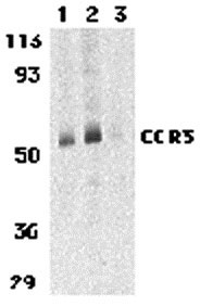
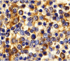
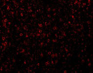
CCR3 Antibody
- 产品详情
- 实验流程
- 背景知识
Application
| WB, IF, E, IHC-P |
|---|---|
| Primary Accession | P51677 |
| Other Accession | NP_847899, 30581170 |
| Reactivity | Human, Mouse, Rat |
| Host | Rabbit |
| Clonality | Polyclonal |
| Isotype | IgG |
| Calculated MW | 41044 Da |
| Concentration (mg/ml) | 1 mg/mL |
| Conjugate | Unconjugated |
| Application Notes | CCR3 antibody can be used for the detection of CCR3 by Western blot at 1 - 2 µg/mL. Antibody can also be used for immunohistochemistry starting at 10 µg/mL. For immunofluorescence start at 20 µg/mL. |
| Gene ID | 1232 |
|---|---|
| Other Names | CCR3 Antibody: CKR3, CD193, CMKBR3, CC-CKR-3, C-C chemokine receptor type 3, Eosinophil eotaxin receptor, C-C CKR-3, chemokine (C-C motif) receptor 3 |
| Target/Specificity | CCR3; At least three isoforms of CCR3 are known to exist; this antibody will detect all three isoforms. |
| Reconstitution & Storage | CCR3 antibody can be stored at 4℃ for three months and -20℃, stable for up to one year. As with all antibodies care should be taken to avoid repeated freeze thaw cycles. Antibodies should not be exposed to prolonged high temperatures. |
| Precautions | CCR3 Antibody is for research use only and not for use in diagnostic or therapeutic procedures. |
| Name | CCR3 |
|---|---|
| Synonyms | CMKBR3 |
| Function | Receptor for C-C type chemokine. Binds and responds to a variety of chemokines, including CCL11, CCL26, CCL7, CCL13, RANTES(CCL5) and CCL15 (PubMed:7622448, PubMed:8642344, PubMed:8676064). Subsequently transduces a signal by increasing the intracellular calcium ions level (PubMed:8676064). In addition acts as a possible functional receptor for NARS1 (PubMed:30171954). |
| Cellular Location | Cell membrane; Multi-pass membrane protein |
| Tissue Location | In eosinophils as well as trace amounts in neutrophils and monocytes. |
For Research Use Only. Not For Use In Diagnostic Procedures.
Provided below are standard protocols that you may find useful for product applications.
BACKGROUND
CCR3 Antibody: Human immunodeficiency virus (HIV) and related virus require coreceptors to infect target cells. Some G protein-coupled receptors including CCR5, CXCR4, CCR3, CCR2b, CCR8, GPR15, STRL33, and CX3CR1 in the chemokine receptor family were recently identified as HIV coreceptors. CCR5, CXCR4 and CCR3 are the principal receptors for HIV fusion and entry of target cells. CCR3 facilitates infection by a subset of virus. CCR3 and CCR5 promote efficient infection of microglia, the major target cells in the CNS. High levels of CCR3 and CXCR4 expression were found on the neurons from both the central and peripheral nervous systems. The CCR3 ligand, eotaxin, and an anti-CCR3 antibody inhibited HIV infection of microglia. These results indicate CCR3 plays an important role in HIV infection of CNS.
REFERENCES
Feng Y, Broder CC, Kennedy PE, et al. HIV-1 entry cofactor: functional cDNA cloning of a seven-transmembrane, G protein-coupled receptor. Science 1996; 272:872-7.
Deng H, Liu R, Ellmeier W, et al. Identification of a major co-receptor for primary isolates of HIV-1. Nature 1996; 381:661-6.
Choe H, Farzan M, Sun Y, et al. The β-chemokine receptors CCR3 and CCR5 facilitate infection by primary HIV-1 isolates. Cell 1996; 85:1135-48.
He J, Chen Y, Farzan M, et al. CCR3 and CCR5 are co-receptors for HIV-1 infection of microglia. Nature 1997; 385:645-9.
终于等到您。ABCEPTA(百远生物)抗体产品。
点击下方“我要评价 ”按钮提交您的反馈信息,您的反馈和评价是我们最宝贵的财富之一,
我们将在1-3个工作日内处理您的反馈信息。
如有疑问,联系:0512-88856768 tech-china@abcepta.com.


