


 癌症的基本特征包括细胞增殖、血管生成、迁移、凋亡逃避机制和细胞永生等。找到癌症发生过程中这些通路的关键标记物和对应的抗体用于检测至关重要。
 为您推荐一个泛素化位点预测神器——泛素化分析工具,可以为您的蛋白的泛素化位点作出预测和评分。
 细胞自噬受体图形绘图工具为你的蛋白的细胞受体结合位点作出预测和评分,识别结合到自噬通路中的蛋白是非常重要的,便于让我们理解自噬在正常生理、病理过程中的作用,如发育、细胞分化、神经退化性疾病、压力条件下、感染和癌症。
CX3CL1 Antibody
- 产品详情
- 实验流程
- 背景知识
Application
| WB, IP, E |
|---|---|
| Primary Accession | P78423 |
| Other Accession | AAB50014, 1899259 |
| Reactivity | Human, Mouse |
| Host | Rabbit |
| Clonality | Polyclonal |
| Isotype | IgG |
| Calculated MW | 42203 Da |
| Conjugate | Unconjugated |
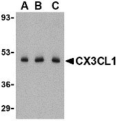
| Application Notes | CX3CL1 antibody can be used for detection of CX3CL1 by Western blot at 0.5 - 2 µg/mL dilution. CX3CL1 is ofen observed migrating at 80 - 100 kDa in SDS-PAGE, presumably due to post-translational modification. |
| Gene ID | 6376 |
|---|---|
| Other Names | CX3CL1 Antibody: NTN, NTT, CXC3, CXC3C, SCYD1, ABCD-3, C3Xkine, fractalkine, neurotactin, FKN, A-152E5.2, Fractalkine, C-X3-C motif chemokine 1, chemokine (C-X3-C motif) ligand 1 |
| Target/Specificity | CX3CL1; |
| Reconstitution & Storage | CX3CL1 antibody can be stored at 4℃ for three months and -20℃, stable for up to one year. As with all antibodies care should be taken to avoid repeated freeze thaw cycles. Antibodies should not be exposed to prolonged high temperatures. |
| Precautions | CX3CL1 Antibody is for research use only and not for use in diagnostic or therapeutic procedures. |
| Name | CX3CL1 {ECO:0000303|PubMed:9024663} |
|---|---|
| Function | Chemokine that acts as a ligand for both CX3CR1 and integrins ITGAV:ITGB3 and ITGA4:ITGB1 (PubMed:12055230, PubMed:21829356, PubMed:23125415, PubMed:9782118, PubMed:9931005). The CX3CR1-CX3CL1 signaling exerts distinct functions in different tissue compartments, such as immune response, inflammation, cell adhesion and chemotaxis (PubMed:12055230, PubMed:9024663, PubMed:9177350, PubMed:9782118). Regulates leukocyte adhesion and migration processes at the endothelium (PubMed:9024663, PubMed:9177350). Can activate integrins in both a CX3CR1-dependent and CX3CR1-independent manner (PubMed:23125415, PubMed:24789099). In the presence of CX3CR1, activates integrins by binding to the classical ligand-binding site (site 1) in integrins (PubMed:23125415, PubMed:24789099). In the absence of CX3CR1, binds to a second site (site 2) in integrins which is distinct from site 1 and enhances the binding of other integrin ligands to site 1 (PubMed:23125415, PubMed:24789099). |
| Cellular Location | Cell membrane; Single-pass type I membrane protein |
| Tissue Location | Expressed in the seminal plasma, endometrial fluid and follicular fluid (at protein level). Small intestine, colon, testis, prostate, heart, brain, lung, skeletal muscle, kidney and pancreas. Most abundant in the brain and heart |
For Research Use Only. Not For Use In Diagnostic Procedures.
Provided below are standard protocols that you may find useful for product applications.
BACKGROUND
CX3CL1 Antibody: Chemokines are a family of proteins associated with the trafficking of leukocytes in immune surveillance and inflammatory cell recruitment. They are classified based on the positions of key cysteine residues. CX3CL1 is a CX3C chemokine known to induce adhesion and migration of leukocytes mediated by a membrane-bound and soluble form respectively. Recent experiments have shown that CX3CL1 can suppress the production of nitrous oxide, interleukin-6, and TNF-α in activated microglia and neuronal cells, suggesting that it may act as an intrinsic inhibitor against neurotoxicity by activated microglia. Its receptor, CX3CR1, also functions as a co-receptor for HIV-1 and HIV-2 envelope fusion and virus infection, which can be inhibited by CX3CL1.
REFERENCES
Bajetto A, Bonavia R, Barbero S, et al. Chemokines and their receptors in the central nervous system. Front. Neuroendocrinol. 2001; 22:147-84.
Umehara H, Goda S, Imai T, et al. Fractalkine, a CX3C-chemokine, functions predominantly as an adhesion molecule in monocytic cell line THP-1. Immunol. Cell. Biol. 2001; 79:298-302.
Mizuno T, Kawanokuchi J, Numata K, et al. Production and neuroprotective functions of fractalkine in the central nervous system. Brain Res. 2003; 979:65-70.
Combadiere C, Salzwedel K, Smith ED, et al. Identification of CX3CR1. A chemotactic receptor for the human CX3C chemokine fractalkine and a fusion coreceptor for HIV-1. J. Biol. Chem.1998; 273:23799-804.
终于等到您。ABCEPTA(百远生物)抗体产品。
点击下方“我要评价 ”按钮提交您的反馈信息,您的反馈和评价是我们最宝贵的财富之一,
我们将在1-3个工作日内处理您的反馈信息。
如有疑问,联系:0512-88856768 tech-china@abcepta.com.


