


 癌症的基本特征包括细胞增殖、血管生成、迁移、凋亡逃避机制和细胞永生等。找到癌症发生过程中这些通路的关键标记物和对应的抗体用于检测至关重要。
 为您推荐一个泛素化位点预测神器——泛素化分析工具,可以为您的蛋白的泛素化位点作出预测和评分。
 细胞自噬受体图形绘图工具为你的蛋白的细胞受体结合位点作出预测和评分,识别结合到自噬通路中的蛋白是非常重要的,便于让我们理解自噬在正常生理、病理过程中的作用,如发育、细胞分化、神经退化性疾病、压力条件下、感染和癌症。
Bnip3L Antibody
- 产品详情
- 实验流程
- 背景知识
Application
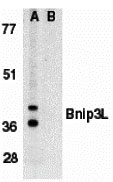
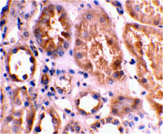
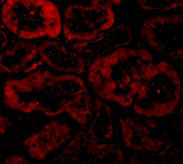
| WB, IF, E, IHC-P |
|---|---|
| Primary Accession | O60238 |
| Other Accession | NP_004322, 4138825 |
| Reactivity | Human |
| Host | Rabbit |
| Clonality | Polyclonal |
| Isotype | IgG |
| Calculated MW | 24 KDa |
| Concentration (mg/ml) | 1 mg/mL |
| Conjugate | Unconjugated |
| Application Notes | Bnip3L antibody can be used for detection of Bnip3L by Western blot at 1 µg/mL. Antibody can also be used for immunohistochemistry starting at 2 µg/mL. For immunofluorescence start at 10 µg/mL. |
| Gene ID | 665 |
|---|---|
| Other Names | Bnip3L Antibody: NIX, BNIP3a, BNIP3A, BNIP3H, NIX, Adenovirus E1B19K-binding protein B5, NIP3L, BCL2/adenovirus E1B 19kDa interacting protein 3-like |
| Target/Specificity | BNIP3L; At least two isoforms of Bnip3L are known to exist. |
| Reconstitution & Storage | Bnip3L antibody can be stored at 4℃ for three months and -20℃, stable for up to one year. As with all antibodies care should be taken to avoid repeated freeze thaw cycles. Antibodies should not be exposed to prolonged high temperatures. |
| Precautions | Bnip3L Antibody is for research use only and not for use in diagnostic or therapeutic procedures. |
For Research Use Only. Not For Use In Diagnostic Procedures.
Provided below are standard protocols that you may find useful for product applications.
BACKGROUND
Bnip3L Antibody: Members in the Bcl-2 family are critical regulators of apoptosis by either inhibiting or promoting cell death. Bcl-2 homology 3 (BH3) domain is a potent death domain. BH3 domain containing pro-apoptotic proteins, including Bad, Bax, Bid, Bik, Hrk, Nip3, and Bim, form a growing subclass of the Bcl-2 family. A novel BH3 domain containing protein was recently identified and designated Bnip3L, Bnip3alpha, and Nix (for Nip3-like protein X). Bnip3L/Bnip3alpha/Nix is a homolog of the E1B 19K/Bcl-2 binding and pro-apoptotic protein Bnip3. Overexpression of Bnip3L induces apoptosis. Bnip3L interacts with and overcomes suppresses by Bcl-2 and Bcl-xL. Bnip3L is localized in mitochondria. The messenger RNA of Bnip3L is ubiquitously expressed in human tissues. Bnip3L and Bnip3 form a new subfamily of the pro-apoptotic mitochondrial proteins.
REFERENCES
Matsushima M, Fujiwara T, Takahashi E, et al. Isolation, mapping, and functional analysis of a novel human cDNA (BNIP3L) encoding a protein homologous to human NIP3. Genes Chromosomes Cancer 1998; 21:230-5
Yasuda M, Han JW, Dionne CA, et al. BNIP3α: a human homolog of mitochondrial proapoptotic protein BNIP3. Cancer Res. 1999; 59:533-7
Chen G, Cizeau J, Vande Velde C, et al. Nix and Nip3 form a subfamily of pro-apoptotic mitochondrial proteins. J. Biol. Chem. 1999; 274:7-10.
Imazu T, Shimizu S, Tagami S, et al. Bcl-2/E1B 19 kDa-interacting protein 3-like protein (Bnip3L) interacts with bcl-2/Bcl-xL and induces apoptosis by altering mitochondrial membrane permeability. Oncogene 1999;18:4523-9.
终于等到您。ABCEPTA(百远生物)抗体产品。
点击下方“我要评价 ”按钮提交您的反馈信息,您的反馈和评价是我们最宝贵的财富之一,
我们将在1-3个工作日内处理您的反馈信息。
如有疑问,联系:0512-88856768 tech-china@abcepta.com.


