


 癌症的基本特征包括细胞增殖、血管生成、迁移、凋亡逃避机制和细胞永生等。找到癌症发生过程中这些通路的关键标记物和对应的抗体用于检测至关重要。
 为您推荐一个泛素化位点预测神器——泛素化分析工具,可以为您的蛋白的泛素化位点作出预测和评分。
 细胞自噬受体图形绘图工具为你的蛋白的细胞受体结合位点作出预测和评分,识别结合到自噬通路中的蛋白是非常重要的,便于让我们理解自噬在正常生理、病理过程中的作用,如发育、细胞分化、神经退化性疾病、压力条件下、感染和癌症。
TAB1 Antibody
- 产品详情
- 实验流程
- 背景知识
Application
| WB, IF, ICC, E |
|---|---|
| Primary Accession | Q15750 |
| Other Accession | NP_006107, 5174703 |
| Reactivity | Human, Mouse |
| Host | Rabbit |
| Clonality | Polyclonal |
| Isotype | IgG |
| Calculated MW | 54644 Da |
| Concentration (mg/ml) | 1 mg/mL |
| Conjugate | Unconjugated |
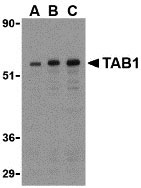
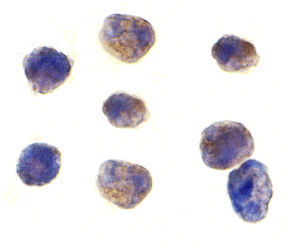
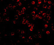
| Application Notes | TAB1 antibody can be used for the detection of TAB1 by Western blot at 0.5 to 2 µg/mL. Antibody can also be used for immunocytochemistry starting at 1 µg/mL. For immunofluorescence start at 2 µg/mL. |
| Gene ID | 10454 |
|---|---|
| Other Names | TAB1 Antibody: 3'-Tab1, MAP3K7IP1, TGF-beta-activated kinase 1 and MAP3K7-binding protein 1, Mitogen-activated protein kinase kinase kinase 7-interacting protein 1, TAK1-binding protein 1, mitogen-activated protein kinase kinase kinase 7 interacting protein 1 |
| Target/Specificity | MAP3K7IP1; |
| Reconstitution & Storage | TAB1 antibody can be stored at 4℃ for three months and -20℃, stable for up to one year. As with all antibodies care should be taken to avoid repeated freeze thaw cycles. Antibodies should not be exposed to prolonged high temperatures. |
| Precautions | TAB1 Antibody is for research use only and not for use in diagnostic or therapeutic procedures. |
| Name | TAB1 |
|---|---|
| Synonyms | MAP3K7IP1 |
| Function | Key adapter protein that plays an essential role in JNK and NF-kappa-B activation and proinflammatory cytokines production in response to stimulation with TLRs and cytokines (PubMed:22307082, PubMed:24403530). Mechanistically, associates with the catalytic domain of MAP3K7/TAK1 to trigger MAP3K7/TAK1 autophosphorylation leading to its full activation (PubMed:10838074, PubMed:25260751, PubMed:37832545). Similarly, associates with MAPK14 and triggers its autophosphorylation and subsequent activation (PubMed:11847341, PubMed:29229647). In turn, MAPK14 phosphorylates TAB1 and inhibits MAP3K7/TAK1 activation in a feedback control mechanism (PubMed:14592977). Also plays a role in recruiting MAPK14 to the TAK1 complex for the phosphorylation of the TAB2 and TAB3 regulatory subunits (PubMed:18021073). |
| Cellular Location | Cytoplasm, cytosol. Endoplasmic reticulum membrane; Peripheral membrane protein; Cytoplasmic side. Note=Recruited to the endoplasmic reticulum following interaction with STING1 |
| Tissue Location | Ubiquitous.. |
For Research Use Only. Not For Use In Diagnostic Procedures.
Provided below are standard protocols that you may find useful for product applications.
BACKGROUND
TAB1 Antibody: TAB1 was identified as a regulator of the MAP kinase kinase kinase TAK1/MAP3K7, which is known to mediate various intracellular signaling pathways, such as those induced by TGF-beta and members of the Toll-IL-1R (TIR) superfamily, thus acting as an intermediate in both proliferative and innate and adaptive immune responses. This protein, together with either TAB2 or TAB3, activates TAK1 kinase in response to upstream signals. It has been shown that the C-terminal portion of TAB1 is sufficient for binding and activation of TAK1, while a portion of the N-terminus acts as a dominant-negative inhibitor of TGF-beta, demonstrating how this protein can function as a mediator between TGF-beta receptors and TAK1.
REFERENCES
Shibuya H, Yamaguchi K, Shirakabe K, et al. TAB1: an activator of the TAK1 MAPKKK in TGF-β signal transduction. Science 1996; 272:1179-82.
Irie T, Muta T, and Takeshige K. TAK1 mediates an activation signal from toll-like receptor(s) to nuclear factor-κB in lipopolysaccharide-stimulated macrophages. FEBS Lett. 2000; 467:160-4.
Akira S and Takeda K. Toll-like receptor Signalling. Nat. Rev. Immunol. 2004; 4:499-511.
Jiang Z, Ninomiya-Tsuji J, Qian Y, et al. Interleukin-1 (IL-1) receptor-associated kinase-dependent IL-1-induced signaling complexes phosphorylate TAK1 and TAB2 at the plasma membrane and activate TAK1 in the cytosol. Mol. Cell Biol. 2002; 22:7158-67.
终于等到您。ABCEPTA(百远生物)抗体产品。
点击下方“我要评价 ”按钮提交您的反馈信息,您的反馈和评价是我们最宝贵的财富之一,
我们将在1-3个工作日内处理您的反馈信息。
如有疑问,联系:0512-88856768 tech-china@abcepta.com.


