


 癌症的基本特征包括细胞增殖、血管生成、迁移、凋亡逃避机制和细胞永生等。找到癌症发生过程中这些通路的关键标记物和对应的抗体用于检测至关重要。
 为您推荐一个泛素化位点预测神器——泛素化分析工具,可以为您的蛋白的泛素化位点作出预测和评分。
 细胞自噬受体图形绘图工具为你的蛋白的细胞受体结合位点作出预测和评分,识别结合到自噬通路中的蛋白是非常重要的,便于让我们理解自噬在正常生理、病理过程中的作用,如发育、细胞分化、神经退化性疾病、压力条件下、感染和癌症。
Bik Antibody
- 产品详情
- 实验流程
- 背景知识
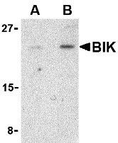
Application
| WB, IF, ICC, E |
|---|---|
| Primary Accession | Q13323 |
| Other Accession | CAG30276, 47678311 |
| Reactivity | Human, Mouse |
| Host | Rabbit |
| Clonality | Polyclonal |
| Isotype | IgG |
| Calculated MW | 18016 Da |
| Concentration (mg/ml) | 1 mg/mL |
| Conjugate | Unconjugated |
| Application Notes | BIK antibody can be used for the detection of BIK by Western blot at 1 - 2 µg/mL. Antibody can also be used for immunocytochemistry starting at 1 µg/mL. For immunofluorescence start at 10 µg/mL. |
| Gene ID | 638 |
|---|---|
| Other Names | Bik Antibody: BP4, NBK, BIP1, Bcl-2-interacting killer, Apoptosis inducer NBK, BCL2-interacting killer (apoptosis-inducing) |
| Target/Specificity | BIK; |
| Reconstitution & Storage | Bik antibody can be stored at 4℃ for three months and -20℃, stable for up to one year. As with all antibodies care should be taken to avoid repeated freeze thaw cycles. Antibodies should not be exposed to prolonged high temperatures. |
| Precautions | Bik Antibody is for research use only and not for use in diagnostic or therapeutic procedures. |
| Name | BIK {ECO:0000303|PubMed:7478623, ECO:0000312|HGNC:HGNC:1051} |
|---|---|
| Function | Accelerates programmed cell death. Association to the apoptosis repressors Bcl-X(L), BHRF1, Bcl-2 or its adenovirus homolog E1B 19k protein suppresses this death-promoting activity. Does not interact with BAX. |
| Cellular Location | Endomembrane system; Single-pass membrane protein. Mitochondrion membrane {ECO:0000250|UniProtKB:O70337}; Single-pass membrane protein. Note=Around the nuclear envelope, and in cytoplasmic membranes. |
For Research Use Only. Not For Use In Diagnostic Procedures.
Provided below are standard protocols that you may find useful for product applications.
BACKGROUND
Bik Antibody: Apoptosis plays a major role in normal organism development, tissue homeostasis, and removal of damaged cells and is caused by the activation of proteolytic enzymes termed caspases. Proteins that comprise the Bcl-2 family appear to control the activation of these enzymes. One such protein BIK was recently identified as an endoplasmic reticulum (ER)-residing pro-apoptotic member of the Bcl-2 homology domain-3 (BH3)-only group of the Bcl-2 family that stimulates mitochondrial release of cytochrome c following p53 induction of apoptosis. A significant fraction of BIK is found as an ER transmembrane protein, with most of the protein facing the cytosol. Restricting BIK to the ER membrane by replacing the transmembrane region with that of the ER-selective membrane anchor of cytochrome b resulted in a decreased cytochrome c release from mitochondria and a corresponding drop in cell death. Recent evidence suggests that BIK cooperates with NOXA, another BH3-only protein, to somehow enhance the activation of Bax to stimulate the rapid release of cytochrome c from mitochondria.
REFERENCES
Lockshin RA, Osborne B, and Zakeri Z. Cell death in the third millennium. Cell Death Differ. 2000; 7:2-7.
Germain M, Mathai JP, and Shore GC. BH-3-only BIK functions at the endoplasmic reticulum to stimulate cytochrome c release from mitochondria. J. Biol. Chem. 277:18053-60.
Germain M, Mathai JP, McBride HM, et al. Endoplasmic reticulum BIK initiates DRP1-regulated remodelling of mitochondrial cristae during apoptosis. EMBO J. 2005; 24:1546-56.
终于等到您。ABCEPTA(百远生物)抗体产品。
点击下方“我要评价 ”按钮提交您的反馈信息,您的反馈和评价是我们最宝贵的财富之一,
我们将在1-3个工作日内处理您的反馈信息。
如有疑问,联系:0512-88856768 tech-china@abcepta.com.


