


 癌症的基本特征包括细胞增殖、血管生成、迁移、凋亡逃避机制和细胞永生等。找到癌症发生过程中这些通路的关键标记物和对应的抗体用于检测至关重要。
 为您推荐一个泛素化位点预测神器——泛素化分析工具,可以为您的蛋白的泛素化位点作出预测和评分。
 细胞自噬受体图形绘图工具为你的蛋白的细胞受体结合位点作出预测和评分,识别结合到自噬通路中的蛋白是非常重要的,便于让我们理解自噬在正常生理、病理过程中的作用,如发育、细胞分化、神经退化性疾病、压力条件下、感染和癌症。
Emerin Antibody

- 产品详情
- 实验流程
- 背景知识
Application
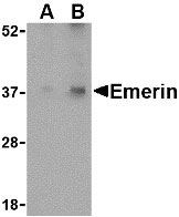
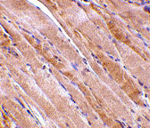
| WB, IF, E, IHC-P |
|---|---|
| Primary Accession | P50402 |
| Other Accession | NP_000108, 4557553 |
| Reactivity | Human, Mouse, Rat |
| Host | Rabbit |
| Clonality | Polyclonal |
| Isotype | IgG |
| Calculated MW | 28994 Da |
| Concentration (mg/ml) | 1 mg/mL |
| Conjugate | Unconjugated |
| Application Notes | Emerin antibody can be used for detection of Emerin by Western blot at 0.5 - 1 µg/mL. Antibody can also be used for immunohistochemistry starting at 2.5 µg/mL. For immunofluorescence start at 10 µg/mL. |
| Gene ID | 2010 |
|---|---|
| Other Names | Emerin Antibody: STA, EDMD, LEMD5, STA, Emerin, emerin |
| Target/Specificity | EMD; |
| Reconstitution & Storage | Emerin antibody can be stored at 4℃ for three months and -20℃, stable for up to one year. As with all antibodies care should be taken to avoid repeated freeze thaw cycles. Antibodies should not be exposed to prolonged high temperatures. |
| Precautions | Emerin Antibody is for research use only and not for use in diagnostic or therapeutic procedures. |
| Name | EMD |
|---|---|
| Synonyms | EDMD, STA |
| Function | Stabilizes and promotes the formation of a nuclear actin cortical network. Stimulates actin polymerization in vitro by binding and stabilizing the pointed end of growing filaments. Inhibits beta- catenin activity by preventing its accumulation in the nucleus. Acts by influencing the nuclear accumulation of beta-catenin through a CRM1- dependent export pathway. Links centrosomes to the nuclear envelope via a microtubule association. Required for proper localization of non- farnesylated prelamin-A/C. Together with NEMP1, contributes to nuclear envelope stiffness in germ cells (PubMed:32923640). EMD and BAF are cooperative cofactors of HIV-1 infection. Association of EMD with the viral DNA requires the presence of BAF and viral integrase. The association of viral DNA with chromatin requires the presence of BAF and EMD. |
| Cellular Location | Nucleus inner membrane; Single-pass membrane protein; Nucleoplasmic side. Nucleus outer membrane. Note=Colocalized with BANF1 at the central region of the assembling nuclear rim, near spindle-attachment sites. The accumulation of different intermediates of prelamin-A/C (non-farnesylated or carboxymethylated farnesylated prelamin-A/C) in fibroblasts modify its localization in the nucleus |
| Tissue Location | Skeletal muscle, heart, colon, testis, ovary and pancreas |
For Research Use Only. Not For Use In Diagnostic Procedures.
Provided below are standard protocols that you may find useful for product applications.
BACKGROUND
Emerin Antibody: Emerin is a serine-rich nuclear membrane protein and a member of the nuclear lamina-associated protein family that includes proteins such as LAP2 and MAN1. Each family member, including Emerin, has an ~40 amino acid LEM-domains that binds barrier-to-autointegration (BANF1), a conserved chromatin protein that also serves as a host cell component of retroviral integration complexes, including that of HIV. Emerin is anchored at the inner membrane of the nuclear envelope where it binds to nuclear intermediate filaments that are formed by lamin proteins. Dreifuss-Emery muscular dystrophy is an X-linked inherited degenerative myopathy resulting from mutation in the emerin gene.
REFERENCES
Schirmer EC, Florens L, Guan T, et al. Nuclear membrane proteins with potential disease links found by subtractive proteomics. Science 2003; 301:1380-2.
Cai M, Huang Y, Ghirlando R, et al. Solution structure of the constant region of nuclear envelope protein LAP2 reveals two LEM-domain structures: one binds BAF and the other binds DNA. EMBO J. 2001; 20:4399-407.
Chen H and Engelman A. The barrier-to-autointegration protein is a host factor for HIV type 1 integration. Proc. Natl. Acad. Sci. USA 1998; 95:15270-4.
Hutchison CJ. Lamins: building blocks or regulators of gene expression? Nat. Rev. Mol. Cell Biol. 2002; 3:848-58.
终于等到您。ABCEPTA(百远生物)抗体产品。
点击下方“我要评价 ”按钮提交您的反馈信息,您的反馈和评价是我们最宝贵的财富之一,
我们将在1-3个工作日内处理您的反馈信息。
如有疑问,联系:0512-88856768 tech-china@abcepta.com.


