


 癌症的基本特征包括细胞增殖、血管生成、迁移、凋亡逃避机制和细胞永生等。找到癌症发生过程中这些通路的关键标记物和对应的抗体用于检测至关重要。
 为您推荐一个泛素化位点预测神器——泛素化分析工具,可以为您的蛋白的泛素化位点作出预测和评分。
 细胞自噬受体图形绘图工具为你的蛋白的细胞受体结合位点作出预测和评分,识别结合到自噬通路中的蛋白是非常重要的,便于让我们理解自噬在正常生理、病理过程中的作用,如发育、细胞分化、神经退化性疾病、压力条件下、感染和癌症。
PDCD5 Antibody
- 产品详情
- 实验流程
- 背景知识
Application
| WB, ICC, E |
|---|---|
| Primary Accession | Q9UEW8 |
| Other Accession | CAG33215, 115430252 |
| Reactivity | Human, Mouse, Rat |
| Host | Rabbit |
| Clonality | Polyclonal |
| Isotype | IgG |
| Calculated MW | 59474 Da |
| Concentration (mg/ml) | 1 mg/mL |
| Conjugate | Unconjugated |
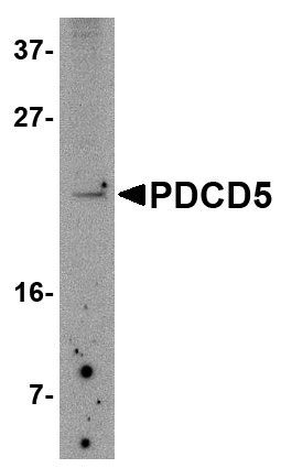
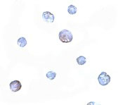
| Application Notes | PDCD5 antibody can be used for detection of PDCD5 by Western blot at 2.5 µg/mL. Antibody can also be used for immunocytochemistry starting at 5 µg/mL. |
| Gene ID | 27347 |
|---|---|
| Other Names | STE20/SPS1-related proline-alanine-rich protein kinase, Ste-20-related kinase, 2.7.11.1, DCHT, Serine/threonine-protein kinase 39, STK39, SPAK |
| Target/Specificity | STK39; |
| Reconstitution & Storage | PDCD5 antibody can be stored at 4℃ for three months and -20℃, stable for up to one year. As with all antibodies care should be taken to avoid repeated freeze thaw cycles. Antibodies should not be exposed to prolonged high temperatures. |
| Precautions | PDCD5 Antibody is for research use only and not for use in diagnostic or therapeutic procedures. |
| Name | STK39 |
|---|---|
| Function | Effector serine/threonine-protein kinase component of the WNK-SPAK/OSR1 kinase cascade, which is involved in various processes, such as ion transport, response to hypertonic stress and blood pressure (PubMed:16669787, PubMed:18270262, PubMed:21321328, PubMed:34289367). Specifically recognizes and binds proteins with a RFXV motif (PubMed:16669787, PubMed:21321328). Acts downstream of WNK kinases (WNK1, WNK2, WNK3 or WNK4): following activation by WNK kinases, catalyzes phosphorylation of ion cotransporters, such as SLC12A1/NKCC2, SLC12A2/NKCC1, SLC12A3/NCC, SLC12A5/KCC2 or SLC12A6/KCC3, regulating their activity (PubMed:21321328). Mediates regulatory volume increase in response to hyperosmotic stress by catalyzing phosphorylation of ion cotransporters SLC12A1/NKCC2, SLC12A2/NKCC1 and SLC12A6/KCC3 downstream of WNK1 and WNK3 kinases (PubMed:12740379, PubMed:16669787, PubMed:21321328). Phosphorylation of Na-K-Cl cotransporters SLC12A2/NKCC1 and SLC12A2/NKCC1 promote their activation and ion influx; simultaneously, phosphorylation of K-Cl cotransporters SLC12A5/KCC2 and SLC12A6/KCC3 inhibit their activity, blocking ion efflux (PubMed:16669787, PubMed:19665974, PubMed:21321328). Acts as a regulator of NaCl reabsorption in the distal nephron by mediating phosphorylation and activation of the thiazide-sensitive Na-Cl cotransporter SLC12A3/NCC in distal convoluted tubule cells of kidney downstream of WNK4 (PubMed:18270262). Mediates the inhibition of SLC4A4, SLC26A6 as well as CFTR activities (By similarity). Phosphorylates RELT (By similarity). |
| Cellular Location | Cytoplasm. Nucleus. Note=Nucleus when caspase-cleaved. |
| Tissue Location | Predominantly expressed in brain and pancreas followed by heart, lung, kidney, skeletal muscle, liver, placenta and testis. |
For Research Use Only. Not For Use In Diagnostic Procedures.
Provided below are standard protocols that you may find useful for product applications.
BACKGROUND
PDCD5 Antibody: Programmed cell death 5 (PDCD5), a human apoptosis-related protein, is thought to play an early and universal role in apoptosis. PDCD5 is widely expressed and is upregulated in cells undergoing apoptosis, where it translocates rapidly from the cytoplasm to the nucleus. PDCD5 has a compact core structure of low flexibility with two mobile alpha-helices at N-terminal and a flexible unstructured C-terminal region. The charged residues are crucial for the ability of apoptosis-promoting and cell translocation of the protein. PDCD5 can facilitate apoptosis and enhance TAJ/TROY-induced paraptosis-like cell death. PDCD5 may play a dual role in the Tip60 pathway. It interacts with Tip60 and functions as a Tip60 co-activator to promote apoptosis. The nucleotide polymorphisms in the 5'-upstream region of PDCD5 affect promoter activity and the susceptibility of a Chinese population to develop chronic myelogenous leukemia and may represent a novel tumor suppressor gene influencing lung cancer.
REFERENCES
Liu H, Wang Y, Zhang Y, et al. TFAR19, a novel apoptosis-related gene cloned from human leukemia cell line TF-1, could enhance apoptosis of some tumor cells induced by growth factor withdrawal. Biochem. Biophys. Res. Commun.1999; 254:203-10.
Chen Y, Sun R, Han W, et al. Nuclear translocation of PDCD5 (TFAR19): an early signal for apoptosis? FEBS Lett.2001; 509:191-6.
Yao H, Xu L, Feng Y, et al. Structure-function correlation of human programmed cell death 5 protein. Arch. Biochem. Biophys.2009 Apr 7.
Wang Y, Li X, Wang L, et al. An alternative form of paraptosis-like cell death triggered by TAJ/TROY and enhanced by PDCD5 overexpression. J. Cell Sci.2004; 117:1525-32.
终于等到您。ABCEPTA(百远生物)抗体产品。
点击下方“我要评价 ”按钮提交您的反馈信息,您的反馈和评价是我们最宝贵的财富之一,
我们将在1-3个工作日内处理您的反馈信息。
如有疑问,联系:0512-88856768 tech-china@abcepta.com.


