


 癌症的基本特征包括细胞增殖、血管生成、迁移、凋亡逃避机制和细胞永生等。找到癌症发生过程中这些通路的关键标记物和对应的抗体用于检测至关重要。
 为您推荐一个泛素化位点预测神器——泛素化分析工具,可以为您的蛋白的泛素化位点作出预测和评分。
 细胞自噬受体图形绘图工具为你的蛋白的细胞受体结合位点作出预测和评分,识别结合到自噬通路中的蛋白是非常重要的,便于让我们理解自噬在正常生理、病理过程中的作用,如发育、细胞分化、神经退化性疾病、压力条件下、感染和癌症。
PIBF1 Antibody
- 产品详情
- 实验流程
- 背景知识
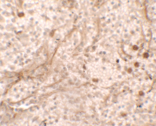
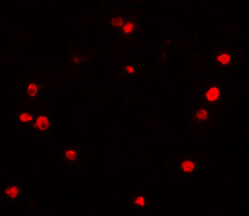
Application
| WB, IF, E, IHC-P |
|---|---|
| Primary Accession | Q8WXW3 |
| Other Accession | NP_006337, 5576958 |
| Reactivity | Human, Mouse, Rat |
| Host | Rabbit |
| Clonality | Polyclonal |
| Isotype | IgG |
| Calculated MW | 89805 Da |
| Concentration (mg/ml) | 1 mg/mL |
| Conjugate | Unconjugated |
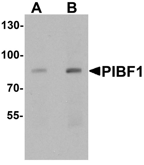
| Application Notes | PIBF1 antibody can be used for detection of PIBF1 by Western blot at 1 - 2 µg/mL. |
| Gene ID | 10464 |
|---|---|
| Other Names | Progesterone-induced-blocking factor 1, PIBF1, C13orf24, PIBF |
| Target/Specificity | PIBF1; Multiple isoforms of PIBF1 exists as a result of alternative splicing event. |
| Reconstitution & Storage | PIBF1 antibody can be stored at 4℃ for three months and -20℃, stable for up to one year. |
| Precautions | PIBF1 Antibody is for research use only and not for use in diagnostic or therapeutic procedures. |
| Name | PIBF1 |
|---|---|
| Synonyms | C13orf24, PIBF |
| Function | Plays a role in ciliogenesis. |
| Cellular Location | Cytoplasm, cytoskeleton, microtubule organizing center, centrosome. Cytoplasm. Secreted Note=In progesterone-treated astrocytoma cells a 57 kDa protein and isoform 1 (90 kDa) have been described, both being located in the intracellular medium and secreted. Respective predominant forms are isoform 1 in the intracellular and the 57 kDa protein in the extracellular medium (PubMed:25218441). [Isoform 4]: Secreted Note=Secreted by progesterone-treated lymphocytes (PubMed:14634107) |
| Tissue Location | Expressed at highest levels in testis. Moderate expression is detected in spleen, thymus, prostate, ovary, small intestine, and colon (PubMed:11935316). Expressed in the first trimester pregnancy decidua (PubMed:12516630). Localized to extravillous cytotrophoblast (at protein level). Also found in syncytiotrophoblast and part of the villous cytotrophoblast. Isoform 1 is expressed in first trimester and term villous trophoblast; trophoblast cells can additionally express other isoforms (PubMed:18817979). Overexpressed in solid tumors from stomach and uterus and in cells from ovary, cervical, breast, lymphoma and leukemia cancer (PubMed:25218441). |
For Research Use Only. Not For Use In Diagnostic Procedures.
Provided below are standard protocols that you may find useful for product applications.
BACKGROUND
PIBF1 Antibody: PIBF1 is synthesized during pregnancy in response to progesterone by T lymphocytes. PIBF1 inhibits arachidonic acid release, controls NK activity, and modifies the cytokine balance exerting an anti-abortive effect. It contains a leucine zipper motif, a basic zipper sequence, a PEST sequence, a nuclear localization signal, an ER membrane retention signal and N-glycosylation and phosphorylation sites. PIBF1 is significantly higher in healthy pregnant women than in women at risk for premature pregnancy termination. Full-length PIBF1 is associated with the nucleus and functions as a transcription factor, whereas secretion of shorter forms which may act as cytokines is induced by activation of the cell.
REFERENCES
Laskarin G, Tokmadzic VS, Strbo N, et al. Progesterone induced blocking factor (PIBF) mediates progesterone induced suppression of decidual lymphocyte cytotoxicity. Am. J. Reprod. Immunol. 2002; 48:201-9.
Lachmann M, Gelbmann D, Kalman E, et al. PIBF (progesterone induced blocking factor) is overexpressed in highly proliferating cells and associated with the centrosome. Int. J. Cancer. 2004; 112:51-60
Polgar B, Kispal G, Lachmann M, et al. Molecular cloning and immunologic characterization of a novel cDNA coding for progesterone-induced blocking factor. J. Immunol. 2003; 171:5956-63.
Raghupathy R, Al-Mutawa E, Al-Azemi M, et al. Progesterone-induced blocking factor (PIBF) modulates cytokine production by lymphocytes from women with recurrent miscarriage or preterm delivery. J. Reprod. Immunol. 2009; 80:91-9.
终于等到您。ABCEPTA(百远生物)抗体产品。
点击下方“我要评价 ”按钮提交您的反馈信息,您的反馈和评价是我们最宝贵的财富之一,
我们将在1-3个工作日内处理您的反馈信息。
如有疑问,联系:0512-88856768 tech-china@abcepta.com.


