


 癌症的基本特征包括细胞增殖、血管生成、迁移、凋亡逃避机制和细胞永生等。找到癌症发生过程中这些通路的关键标记物和对应的抗体用于检测至关重要。
 为您推荐一个泛素化位点预测神器——泛素化分析工具,可以为您的蛋白的泛素化位点作出预测和评分。
 细胞自噬受体图形绘图工具为你的蛋白的细胞受体结合位点作出预测和评分,识别结合到自噬通路中的蛋白是非常重要的,便于让我们理解自噬在正常生理、病理过程中的作用,如发育、细胞分化、神经退化性疾病、压力条件下、感染和癌症。
ULK3 Antibody
- 产品详情
- 实验流程
- 背景知识
Application
| WB, IF, E, IHC-P |
|---|---|
| Primary Accession | Q6PHR2 |
| Other Accession | NP_001092906, 150456432 |
| Reactivity | Human |
| Host | Rabbit |
| Clonality | Polyclonal |
| Isotype | IgG |
| Calculated MW | 53444 Da |
| Concentration (mg/ml) | 1 mg/mL |
| Conjugate | Unconjugated |
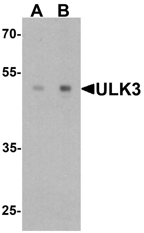
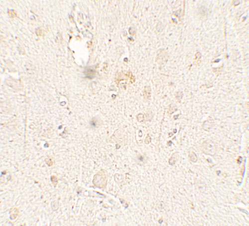
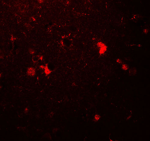
| Application Notes | ULK3 antibody can be used for detection of ULK3 by Western blot at 0.5 - 1 µg/ml. Antibody can also be used for Immunohistochemistry starting at 5 µg/mL. For immunofluorescence start at 20 µg/mL. |
| Gene ID | 25989 |
|---|---|
| Other Names | Serine/threonine-protein kinase ULK3, 2.7.11.1, Unc-51-like kinase 3, ULK3 |
| Target/Specificity | ULK3; ULK3 antibody is human specific. Multiple isoforms of ULK3 are known to exist. |
| Reconstitution & Storage | ULK3 antibody can be stored at 4℃ for three months and -20℃, stable for up to one year. |
| Precautions | ULK3 Antibody is for research use only and not for use in diagnostic or therapeutic procedures. |
| Name | ULK3 |
|---|---|
| Function | Serine/threonine protein kinase that acts as a regulator of Sonic hedgehog (SHH) signaling and autophagy. Acts as a negative regulator of SHH signaling in the absence of SHH ligand: interacts with SUFU, thereby inactivating the protein kinase activity and preventing phosphorylation of GLI proteins (GLI1, GLI2 and/or GLI3). Positively regulates SHH signaling in the presence of SHH: dissociates from SUFU, autophosphorylates and mediates phosphorylation of GLI2, activating it and promoting its nuclear translocation. Phosphorylates in vitro GLI2, as well as GLI1 and GLI3, although less efficiently. Also acts as a regulator of autophagy: following cellular senescence, able to induce autophagy. |
| Cellular Location | Cytoplasm. Note=Localizes to pre-autophagosomal structure during cellular senescence |
| Tissue Location | Widely expressed. Highest levels observed in fetal brain. In adult tissues, high levels in brain, liver and kidney, moderate levels in testis and adrenal gland and low levels in heart, lung, stomach, thymus, prostate and placenta. In the brain, highest expression in the hippocampus, high levels also detected in the cerebellum, olfactory bulb and optic nerve. In the central nervous system, lowest levels in the spinal cord |
For Research Use Only. Not For Use In Diagnostic Procedures.
Provided below are standard protocols that you may find useful for product applications.
BACKGROUND
ULK3 belongs to the Ser/Thr protein kinase superfamily and plays a role in the ATP-dependent phosphorylation of target proteins (1). Knockout of ULK genes results in a severe defect in the autophagy pathway (2). ULK3, like the other Unc-51-like kinases such as ULK1, ULK2 and ULK4, is highly conserved among eukaryotes (3). ULK3 has been shown to be a positive regulator of the Hedgehog signaling pathway by enhancing GLI1 and GLI2 transcriptional activity (4). Furthermore, ULK3 can also interact with SUFU, a protein required for the negative regulation of GLI proteins; this interaction blocks the autophosphorylation of ULK3 and blocks its ability to regulate the GLI proteins (5).
REFERENCES
Suzuki K, Kubota Y, Sekito T, et al. Hierarchy of Atg proteins in pre-autophagosomal structure organization. Genes to Cells 2007; 12:209–18.
Lee EJ and Tournier C. The requirement of uncoordinated 51-like kinase 1 (ULK1) and ULK2 in the regulation of autophagy. Autophagy 2011; 7:689-95.
Zhou X, Babu JR, da Silva S, et al. Unc-51-like kinase 1/2-mediated endocytic processes regulate filopodia extension and branching of sensory axons. Proc. Natl. Acad. Sci. USA 2007; 104:5842-7.
Maloverjan A, Piirsoo M, Michelson P, et al. Identification of a novel serine/threonine kinase ULK3 as a positive regulator of Hedgehog pathway. Exp. Cell Res. 2010; 316:627-37.
终于等到您。ABCEPTA(百远生物)抗体产品。
点击下方“我要评价 ”按钮提交您的反馈信息,您的反馈和评价是我们最宝贵的财富之一,
我们将在1-3个工作日内处理您的反馈信息。
如有疑问,联系:0512-88856768 tech-china@abcepta.com.


