


 癌症的基本特征包括细胞增殖、血管生成、迁移、凋亡逃避机制和细胞永生等。找到癌症发生过程中这些通路的关键标记物和对应的抗体用于检测至关重要。
 为您推荐一个泛素化位点预测神器——泛素化分析工具,可以为您的蛋白的泛素化位点作出预测和评分。
 细胞自噬受体图形绘图工具为你的蛋白的细胞受体结合位点作出预测和评分,识别结合到自噬通路中的蛋白是非常重要的,便于让我们理解自噬在正常生理、病理过程中的作用,如发育、细胞分化、神经退化性疾病、压力条件下、感染和癌症。
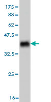
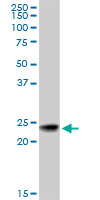
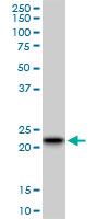
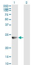
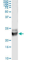
RAB7B Antibody (monoclonal) (M01)
Mouse monoclonal antibody raised against a partial recombinant RAB7B.
- 产品详情
- 实验流程
Application
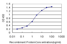
| WB, IP, E |
|---|---|
| Primary Accession | Q96AH8 |
| Other Accession | NM_177403 |
| Reactivity | Human |
| Host | mouse |
| Clonality | monoclonal |
| Isotype | IgG2a Kappa |
| Clone Names | 3B3 |
| Calculated MW | 22511 Da |
| Gene ID | 338382 |
|---|---|
| Other Names | Ras-related protein Rab-7b, RAB7B |
| Target/Specificity | RAB7B (NP_796377, 100 a.a. ~ 199 a.a) partial recombinant protein with GST tag. MW of the GST tag alone is 26 KDa. |
| Dilution | WB~~1:500~1000 IP~~N/A E~~N/A |
| Format | Clear, colorless solution in phosphate buffered saline, pH 7.2 . |
| Storage | Store at -20°C or lower. Aliquot to avoid repeated freezing and thawing. |
| Precautions | RAB7B Antibody (monoclonal) (M01) is for research use only and not for use in diagnostic or therapeutic procedures. |
For Research Use Only. Not For Use In Diagnostic Procedures.
Provided below are standard protocols that you may find useful for product applications.
REFERENCES
1.Clathrin-dependent mechanisms modulate the subcellular distribution of class C Vps/HOPS tether subunits in polarized and nonpolarized cells.Zlatic SA, Tornieri K, L'hernault SW, Faundez V.Mol Biol Cell. 2011 May;22(10):1699-715. Epub 2011 Mar 16.2.Abnormal localization of leucine-rich repeat kinase 2 to the endosomal-lysosomal compartment in lewy body disease.Higashi S, Moore DJ, Yamamoto R, Minegishi M, Sato K, Togo T, Katsuse O, Uchikado H, Furukawa Y, Hino H, Kosaka K, Emson PC, Wada K, Dawson VL, Dawson TM, Arai H, Iseki E.J Neuropathol Exp Neurol. 2009 Sep;68(9):994-1005.3.GIGYF2 is present in endosomal compartments in the mammalian brains and enhances IGF-1-induced ERK1/2 activation.Higashi S, Iseki E, Minegishi M, Togo T, Kabuta T, Wada K.J Neurochem. 2010 Jul 28. [Epub ahead of print]4.SPE-39 Family Proteins Interact with the HOPS Complex and Function in Lysosomal Delivery.Zhu GD, Salazar G, Zlatic SA, Fiza B, Doucette MM, Heilman CJ, Levey AI, Faundez V, L'hernault SW.Mol Biol Cell. 2009 Feb;20(4):1223-1240. Epub 2008 Dec 24.
终于等到您。ABCEPTA(百远生物)抗体产品。
点击下方“我要评价 ”按钮提交您的反馈信息,您的反馈和评价是我们最宝贵的财富之一,
我们将在1-3个工作日内处理您的反馈信息。
如有疑问,联系:0512-88856768 tech-china@abcepta.com.


