


 癌症的基本特征包括细胞增殖、血管生成、迁移、凋亡逃避机制和细胞永生等。找到癌症发生过程中这些通路的关键标记物和对应的抗体用于检测至关重要。
 为您推荐一个泛素化位点预测神器——泛素化分析工具,可以为您的蛋白的泛素化位点作出预测和评分。
 细胞自噬受体图形绘图工具为你的蛋白的细胞受体结合位点作出预测和评分,识别结合到自噬通路中的蛋白是非常重要的,便于让我们理解自噬在正常生理、病理过程中的作用,如发育、细胞分化、神经退化性疾病、压力条件下、感染和癌症。
RPL4 Antibody (monoclonal) (M01)
Mouse monoclonal antibody raised against a partial recombinant RPL4.

- 产品详情
- 实验流程
- 背景知识
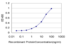
Application
| WB, IF, E |
|---|---|
| Primary Accession | P36578 |
| Other Accession | NM_000968 |
| Reactivity | Human, Mouse, Rat |
| Host | mouse |
| Clonality | monoclonal |
| Isotype | IgG2a Kappa |
| Clone Names | 4A3 |
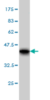
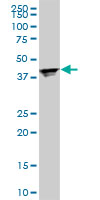
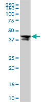
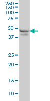
| Calculated MW | 47697 Da |
| Gene ID | 6124 |
|---|---|
| Other Names | 60S ribosomal protein L4, 60S ribosomal protein L1, RPL4, RPL1 |
| Target/Specificity | RPL4 (NP_000959, 251 a.a. ~ 350 a.a) partial recombinant protein with GST tag. MW of the GST tag alone is 26 KDa. |
| Dilution | WB~~1:500~1000 IF~~1:50~200 E~~N/A |
| Format | Clear, colorless solution in phosphate buffered saline, pH 7.2 . |
| Storage | Store at -20°C or lower. Aliquot to avoid repeated freezing and thawing. |
| Precautions | RPL4 Antibody (monoclonal) (M01) is for research use only and not for use in diagnostic or therapeutic procedures. |
For Research Use Only. Not For Use In Diagnostic Procedures.
Provided below are standard protocols that you may find useful for product applications.
BACKGROUND
Ribosomes, the organelles that catalyze protein synthesis, consist of a small 40S subunit and a large 60S subunit. Together these subunits are composed of 4 RNA species and approximately 80 structurally distinct proteins. This gene encodes a ribosomal protein that is a component of the 60S subunit. The protein belongs to the L4E family of ribosomal proteins. It is located in the cytoplasm. As is typical for genes encoding ribosomal proteins, there are multiple processed pseudogenes of this gene dispersed through the genome.
REFERENCES
1.Transmembrane and coiled-coil domain family 1 is a novel protein of the endoplasmic reticulum.Zhang C, Kho YS, Wang Z, Chiang YT, Ng GK, Shaw PC, Wang Y, Qi RZPLoS One. 2014 Jan 14;9(1):e85206. doi: 10.1371/journal.pone.0085206. eCollection 2014 Jan 14.2.Shwachman-Bodian Diamond syndrome is a multi-functional protein implicated in cellular stress responses.Ball HL, Zhang B, Riches JJ, Gandhi R, Li J, Rommens JM, Myers JS.Hum Mol Genet. 2009 Oct 1;18(19):3684-95. Epub 2009 Jul 14.
终于等到您。ABCEPTA(百远生物)抗体产品。
点击下方“我要评价 ”按钮提交您的反馈信息,您的反馈和评价是我们最宝贵的财富之一,
我们将在1-3个工作日内处理您的反馈信息。
如有疑问,联系:0512-88856768 tech-china@abcepta.com.


