


 癌症的基本特征包括细胞增殖、血管生成、迁移、凋亡逃避机制和细胞永生等。找到癌症发生过程中这些通路的关键标记物和对应的抗体用于检测至关重要。
 为您推荐一个泛素化位点预测神器——泛素化分析工具,可以为您的蛋白的泛素化位点作出预测和评分。
 细胞自噬受体图形绘图工具为你的蛋白的细胞受体结合位点作出预测和评分,识别结合到自噬通路中的蛋白是非常重要的,便于让我们理解自噬在正常生理、病理过程中的作用,如发育、细胞分化、神经退化性疾病、压力条件下、感染和癌症。
PACSIN2 Antibody (C-term)
Purified Rabbit Polyclonal Antibody (Pab)
- 产品详情
- 文献引用 : 1
- 实验流程
- 背景知识
Application
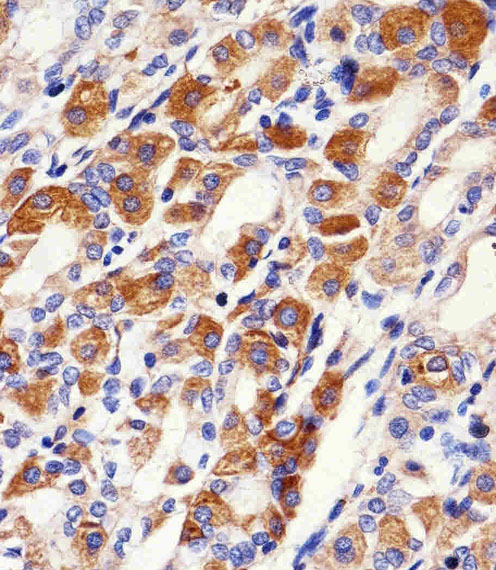
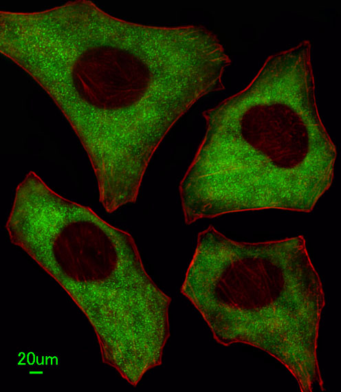
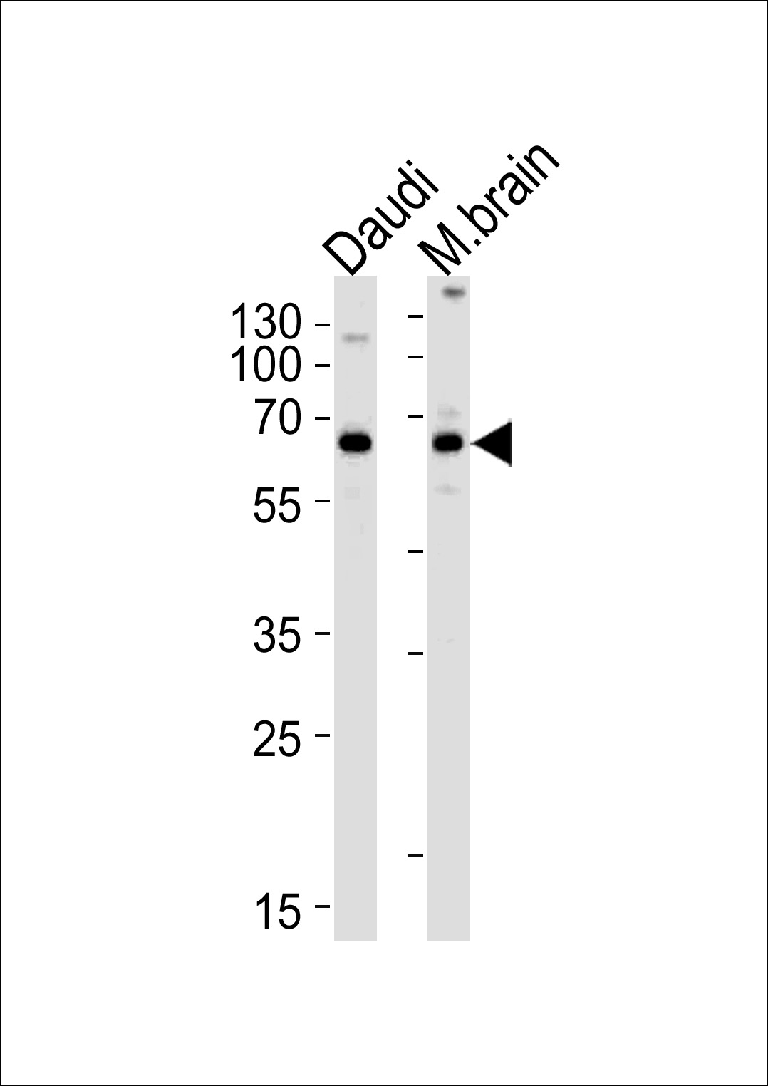
| WB, IF, IHC-P |
|---|---|
| Primary Accession | Q9UNF0 |
| Reactivity | Human, Rat |
| Predicted | Mouse |
| Host | Rabbit |
| Clonality | Polyclonal |
| Calculated MW | 55739 Da |
| Isotype | Rabbit IgG |
| Antigen Source | HUMAN |
| Gene ID | 11252 |
|---|---|
| Antigen Region | 342-371 aa |
| Other Names | PACSIN2; Protein kinase C and casein kinase substrate in neurons protein 2 |
| Dilution | WB~~1:1000 IF~~1:100 IHC-P~~1:100 |
| Target/Specificity | This PACSIN2 antibody is generated from rabbits immunized with a KLH conjugated synthetic peptide between 342-371 amino acids from the C-terminal region of human PACSIN2. |
| Format | Purified polyclonal antibody supplied in PBS with 0.09% (W/V) sodium azide. This antibody is prepared by Saturated Ammonium Sulfate (SAS) precipitation followed by dialysis against PBS. |
| Storage | Maintain refrigerated at 2-8°C for up to 2 weeks. For long term storage store at -20°C in small aliquots to prevent freeze-thaw cycles. |
| Precautions | PACSIN2 Antibody (C-term) is for research use only and not for use in diagnostic or therapeutic procedures. |
| Name | PACSIN2 |
|---|---|
| Function | Regulates the morphogenesis and endocytosis of caveolae (By similarity). Lipid-binding protein that is able to promote the tubulation of the phosphatidic acid-containing membranes it preferentially binds. Plays a role in intracellular vesicle-mediated transport. Involved in the endocytosis of cell-surface receptors like the EGF receptor, contributing to its internalization in the absence of EGF stimulus (PubMed:21693584, PubMed:23129763, PubMed:23236520, PubMed:23596323). Essential for endothelial organization in sprouting angiogenesis, modulates CDH5-based junctions. Facilitates endothelial front-rear polarity during migration by recruiting EHD4 and MICALL1 to asymmetric adherens junctions between leader and follower cells (By similarity). |
| Cellular Location | Cytoplasm {ECO:0000250|UniProtKB:Q9WVE8}. Cytoplasm, cytoskeleton {ECO:0000250|UniProtKB:Q9WVE8}. Cytoplasmic vesicle membrane {ECO:0000250|UniProtKB:Q9WVE8}; Peripheral membrane protein {ECO:0000250|UniProtKB:Q9WVE8}; Cytoplasmic side {ECO:0000250|UniProtKB:Q9WVE8}. Cell projection, ruffle membrane {ECO:0000250|UniProtKB:Q9WVE8}; Peripheral membrane protein {ECO:0000250|UniProtKB:Q9WVE8}; Cytoplasmic side {ECO:0000250|UniProtKB:Q9WVE8}. Early endosome {ECO:0000250|UniProtKB:Q9WVE8}. Recycling endosome membrane. Cell membrane {ECO:0000250|UniProtKB:Q9WVE8}; Peripheral membrane protein {ECO:0000250|UniProtKB:Q9WVE8}; Cytoplasmic side {ECO:0000250|UniProtKB:Q9WVE8}. Cell projection. Membrane, caveola. Cell junction, adherens junction {ECO:0000250|UniProtKB:Q9WVE8}. Note=Detected at the neck of flask- shaped caveolae. Localization to tubular recycling endosomes probably requires interaction with MICALL1 and EHD1 {ECO:0000250|UniProtKB:Q9WVE8} |
| Tissue Location | Widely expressed. |
For Research Use Only. Not For Use In Diagnostic Procedures.

Provided below are standard protocols that you may find useful for product applications.
BACKGROUND
PACSIN may play a role in vesicle formation and transport. This protein homo- and hetero-aggregates with other PACSINs. It also binds dynamin 1, synaptojanin, synapsin 1 and the neural Wiskott-Aldrich syndrome protein (N-WASP). The protein exhibits a cvesicle-like cytoplasmic distribution and is ubiquitously expressed. PACSIN is phosphorylated by casein kinase 2 (CK2) and protein kinase C (PKC). The protein contains 1 FCH domain and 1 SH3 domain.
REFERENCES
Strausberg, R.L., et al., Proc. Natl. Acad. Sci. U.S.A. 99(26):16899-16903 (2002). Wiemann, S., et al., Genome Res. 11(3):422-435 (2001). Ritter, B., et al., FEBS Lett. 454(3):356-362 (1999). Dunham, I., et al., Nature 402(6761):489-495 (1999).
终于等到您。ABCEPTA(百远生物)抗体产品。
点击下方“我要评价 ”按钮提交您的反馈信息,您的反馈和评价是我们最宝贵的财富之一,
我们将在1-3个工作日内处理您的反馈信息。
如有疑问,联系:0512-88856768 tech-china@abcepta.com.


