


 癌症的基本特征包括细胞增殖、血管生成、迁移、凋亡逃避机制和细胞永生等。找到癌症发生过程中这些通路的关键标记物和对应的抗体用于检测至关重要。
 为您推荐一个泛素化位点预测神器——泛素化分析工具,可以为您的蛋白的泛素化位点作出预测和评分。
 细胞自噬受体图形绘图工具为你的蛋白的细胞受体结合位点作出预测和评分,识别结合到自噬通路中的蛋白是非常重要的,便于让我们理解自噬在正常生理、病理过程中的作用,如发育、细胞分化、神经退化性疾病、压力条件下、感染和癌症。
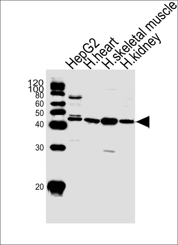
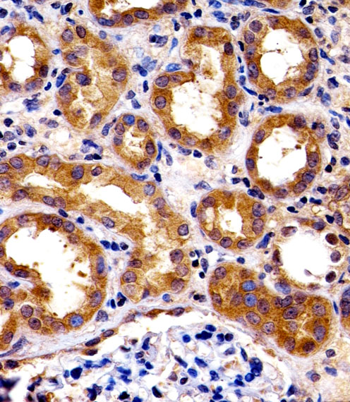
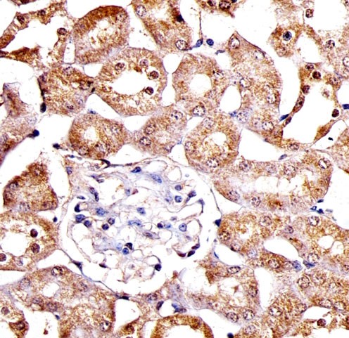
SPHK1 Antibody (N-term P74)
Purified Rabbit Polyclonal Antibody (Pab)
- 产品详情
- 实验流程
- 背景知识
Application
| IHC-P, WB |
|---|---|
| Primary Accession | Q9NYA1 |
| Reactivity | Human |
| Host | Rabbit |
| Clonality | Polyclonal |
| Calculated MW | 42518 Da |
| Isotype | Rabbit IgG |
| Antigen Source | HUMAN |
| Gene ID | 8877 |
|---|---|
| Antigen Region | 59-89 aa |
| Other Names | SPHK1; SPHK; SPK; Sphingosine kinase 1 |
| Dilution | IHC-P~~1:100 WB~~1:1000 |
| Target/Specificity | This SPHK1 antibody is generated from rabbits immunized with a KLH conjugated synthetic peptide between 59-89 amino acids from the N-terminal region of human SPHK1. |
| Format | Purified polyclonal antibody supplied in PBS with 0.09% (W/V) sodium azide. This antibody is prepared by Saturated Ammonium Sulfate (SAS) precipitation followed by dialysis against PBS. |
| Storage | Maintain refrigerated at 2-8°C for up to 2 weeks. For long term storage store at -20°C in small aliquots to prevent freeze-thaw cycles. |
| Precautions | SPHK1 Antibody (N-term P74) is for research use only and not for use in diagnostic or therapeutic procedures. |
| Name | SPHK1 (HGNC:11240) |
|---|---|
| Function | Catalyzes the phosphorylation of sphingosine to form sphingosine 1-phosphate (SPP), a lipid mediator with both intra- and extracellular functions. Also acts on D-erythro-sphingosine and to a lesser extent sphinganine, but not other lipids, such as D,L-threo- dihydrosphingosine, N,N-dimethylsphingosine, diacylglycerol, ceramide, or phosphatidylinositol (PubMed:11923095, PubMed:20577214, PubMed:23602659, PubMed:24929359, PubMed:29662056). In contrast to proapoptotic SPHK2, has a negative effect on intracellular ceramide levels, enhances cell growth and inhibits apoptosis (PubMed:16118219). Involved in the regulation of inflammatory response and neuroinflammation. Via the product sphingosine 1-phosphate, stimulates TRAF2 E3 ubiquitin ligase activity, and promotes activation of NF- kappa-B in response to TNF signaling leading to IL17 secretion (PubMed:20577214). In response to TNF and in parallel to NF-kappa-B activation, negatively regulates RANTES induction through p38 MAPK signaling pathway (PubMed:23935096). Involved in endocytic membrane trafficking induced by sphingosine, recruited to dilate endosomes, also plays a role on later stages of endosomal maturation and membrane fusion independently of its kinase activity (PubMed:24929359, PubMed:28049734). In Purkinje cells, seems to be also involved in the regulation of autophagosome-lysosome fusion upon VEGFA (PubMed:25417698). |
| Cellular Location | Cytoplasm. Nucleus. Cell membrane. Endosome membrane; Peripheral membrane protein. Membrane, clathrin-coated pit. Synapse {ECO:0000250|UniProtKB:Q8CI15} Note=Translocated from the cytoplasm to the plasma membrane in a CIB1- dependent manner (PubMed:19854831). Binds to membranes containing negatively charged lipids but not neutral lipids (PubMed:24929359) Recruited to endocytic membranes by sphingosine where promotes membrane fusion (By similarity). {ECO:0000250|UniProtKB:Q8CI15, ECO:0000269|PubMed:19854831, ECO:0000269|PubMed:24929359} |
| Tissue Location | Widely expressed with highest levels in adult liver, kidney, heart and skeletal muscle. Expressed in brain cortex (at protein level) (PubMed:29662056). |
For Research Use Only. Not For Use In Diagnostic Procedures.
Provided below are standard protocols that you may find useful for product applications.
BACKGROUND
Sphingosine Kinase (SphK) catalyzes the phosphorylation of the lipid sphingosine, creating the bioactive lipid sphingosine-1-phosphate (S1P). S1P subsequently signals through cell surface G protein-coupled receptors, as well as intracellularly, to modulate cell proliferation, survival, motility and differentiation. SphK is an important signaling enzyme which is activated by diverse agents, including growth factors that signal through receptor tyrosine kinases, agents activating G protein-coupled receptors, and immunoglobulin receptors. Two SphK isotypes, SphK-1 and SphK-2, have been cloned, and both isotypes are ubiquitously expressed. SphK-1 has been shown to mediate cell growth, prevention of apoptosis, and cellular transformation, and is upregulated in a variety of human tumors. In contrast, SphK-2 increases apoptosis, and may be responsible for phosphorylating and activating the immunosuppressive drug FTY720.
REFERENCES
Ota, T., et al., Nat. Genet. 36(1):40-45 (2004).
Nava, V.E., et al., FEBS Lett. 473(1):81-84 (2000).
Melendez, A.J., et al., Gene 251(1):19-26 (2000).
Pitson, S.M., et al., Biochem. J. 350 Pt 2, 429-441 (2000).
终于等到您。ABCEPTA(百远生物)抗体产品。
点击下方“我要评价 ”按钮提交您的反馈信息,您的反馈和评价是我们最宝贵的财富之一,
我们将在1-3个工作日内处理您的反馈信息。
如有疑问,联系:0512-88856768 tech-china@abcepta.com.


