


 癌症的基本特征包括细胞增殖、血管生成、迁移、凋亡逃避机制和细胞永生等。找到癌症发生过程中这些通路的关键标记物和对应的抗体用于检测至关重要。
 为您推荐一个泛素化位点预测神器——泛素化分析工具,可以为您的蛋白的泛素化位点作出预测和评分。
 细胞自噬受体图形绘图工具为你的蛋白的细胞受体结合位点作出预测和评分,识别结合到自噬通路中的蛋白是非常重要的,便于让我们理解自噬在正常生理、病理过程中的作用,如发育、细胞分化、神经退化性疾病、压力条件下、感染和癌症。
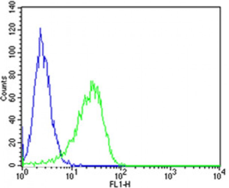
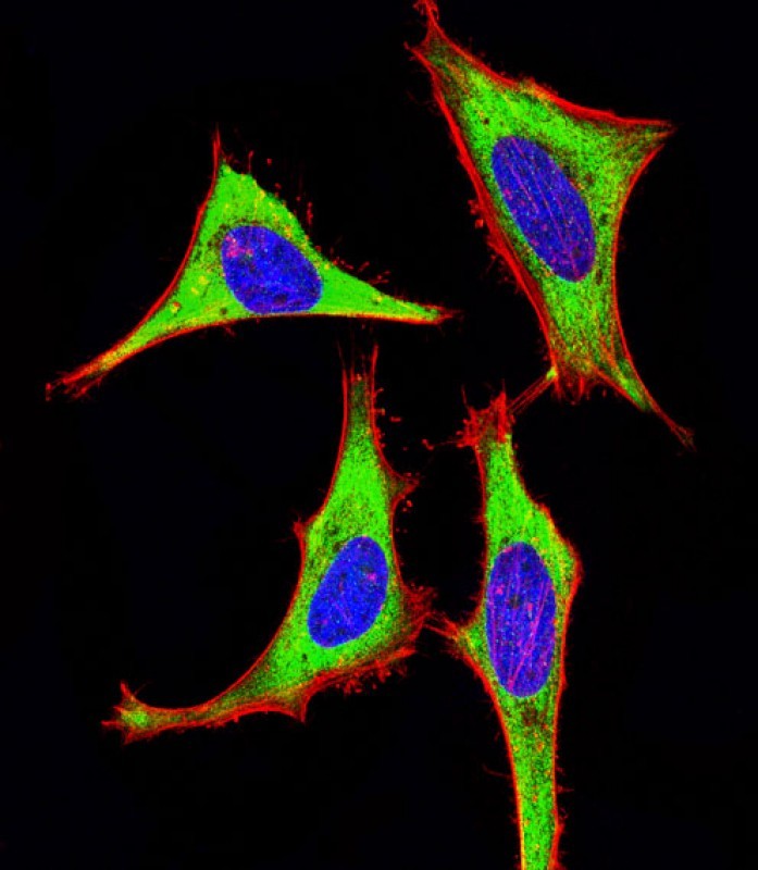
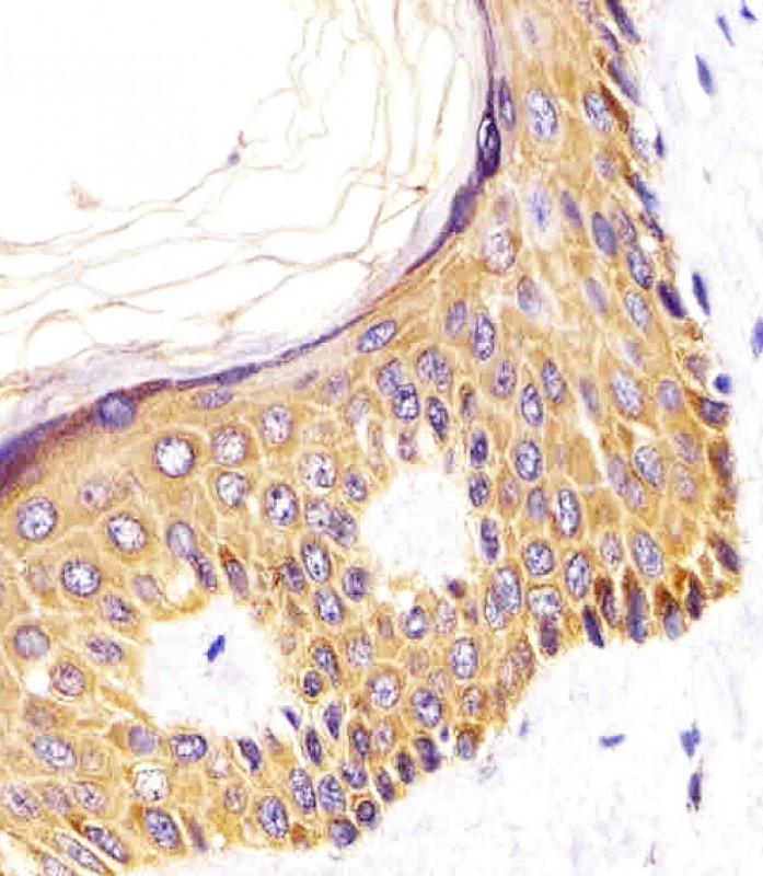
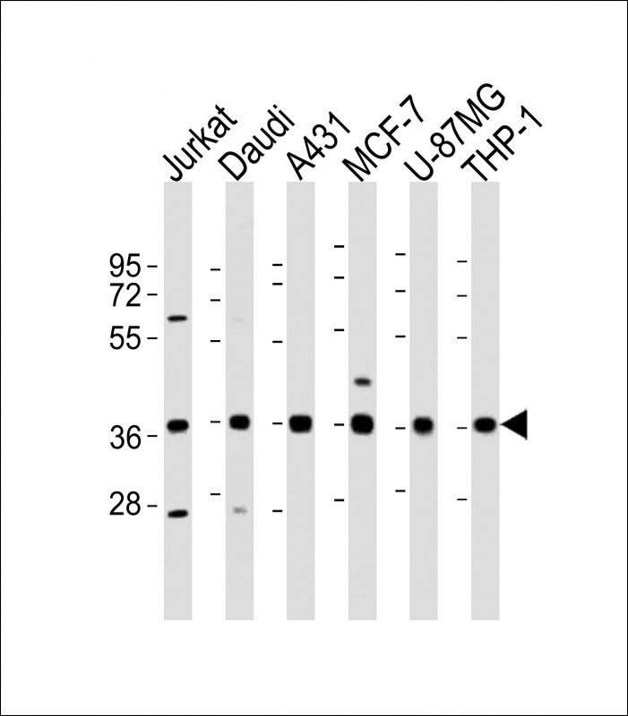
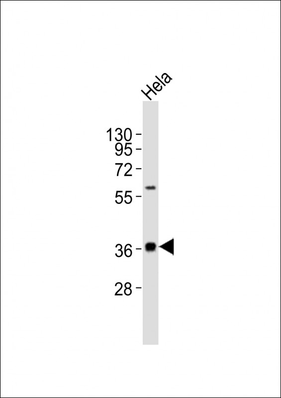
MICA Antibody (Center)
Affinity Purified Rabbit Polyclonal Antibody (Pab)
- 产品详情
- 实验流程
- 背景知识
Application
| WB, IHC-P, FC, IF |
|---|---|
| Primary Accession | Q29983 |
| Reactivity | Human |
| Host | Rabbit |
| Clonality | Polyclonal |
| Calculated MW | 42915 Da |
| Isotype | Rabbit IgG |
| Antigen Source | HUMAN |
| Gene ID | 100507436 |
|---|---|
| Antigen Region | 68-97 aa |
| Other Names | MHC class I polypeptide-related sequence A, MIC-A, MICA {ECO:0000312|EMBL:CAI419071} |
| Dilution | WB~~1:1000 IHC-P~~1:100~500 FC~~1:25 IF~~1:25 |
| Target/Specificity | This MICA antibody is generated from rabbits immunized with a KLH conjugated synthetic peptide between 68-97 amino acids from the Central region of human MICA. |
| Format | Purified polyclonal antibody supplied in PBS with 0.09% (W/V) sodium azide. This antibody is purified through a protein A column, followed by peptide affinity purification. |
| Storage | Maintain refrigerated at 2-8°C for up to 2 weeks. For long term storage store at -20°C in small aliquots to prevent freeze-thaw cycles. |
| Precautions | MICA Antibody (Center) is for research use only and not for use in diagnostic or therapeutic procedures. |
| Name | MICA {ECO:0000312|EMBL:CAI41907.1} |
|---|---|
| Function | Widely expressed membrane-bound protein which acts as a ligand to stimulate an activating receptor KLRK1/NKG2D, expressed on the surface of essentially all human natural killer (NK), gammadelta T and CD8 alphabeta T-cells (PubMed:11491531, PubMed:11777960). Up- regulated in stressed conditions, such as viral and bacterial infections or DNA damage response, serves as signal of cellular stress, and engagement of KLRK1/NKG2D by MICA triggers NK-cells resulting in a range of immune effector functions, such as cytotoxicity and cytokine production (PubMed:10426993). |
| Cellular Location | Cell membrane; Single-pass type I membrane protein. Cytoplasm Note=Expressed on the cell surface in gastric epithelium, endothelial cells and fibroblasts and in the cytoplasm in keratinocytes and monocytes. Infection with human adenovirus 5 suppresses cell surface expression due to the adenoviral E3-19K protein which causes retention in the endoplasmic reticulum. |
| Tissue Location | Widely expressed with the exception of the central nervous system where it is absent. Expressed predominantly in gastric epithelium and also in monocytes, keratinocytes, endothelial cells, fibroblasts and in the outer layer of Hassal's corpuscles within the medulla of normal thymus. In skin, expressed mainly in the keratin layers, basal cells, ducts and follicles. Also expressed in many, but not all, epithelial tumors of lung, breast, kidney, ovary, prostate and colon. In thyomas, overexpressed in cortical and medullar epithelial cells. Tumors expressing MICA display increased levels of gamma delta T-cells. |
For Research Use Only. Not For Use In Diagnostic Procedures.
Provided below are standard protocols that you may find useful for product applications.
BACKGROUND
MICA is the higly polymorphic MHC (HLA) class I chain-related gene A. The protein product is expressed on the cell surface, although unlike canonical class I molecules does not seem to associate with beta-2-microglobulin. It is thought that MICA functions as a stress-induced antigen that is broadly recognized by intestinal epithelial gamma delta T cells.
REFERENCES
Bahram,S., et.al., Proc. Natl. Acad. Sci. U.S.A. 91 (14), 6259-6263 (1994) Klein,J.et.al., Proc. Natl. Acad. Sci. U.S.A. 91 (14), 6251-6252 (1994) Parham,P., et.al., J. Immunol. 142 (11), 3937-3950 (1989)
终于等到您。ABCEPTA(百远生物)抗体产品。
点击下方“我要评价 ”按钮提交您的反馈信息,您的反馈和评价是我们最宝贵的财富之一,
我们将在1-3个工作日内处理您的反馈信息。
如有疑问,联系:0512-88856768 tech-china@abcepta.com.


