


 癌症的基本特征包括细胞增殖、血管生成、迁移、凋亡逃避机制和细胞永生等。找到癌症发生过程中这些通路的关键标记物和对应的抗体用于检测至关重要。
 为您推荐一个泛素化位点预测神器——泛素化分析工具,可以为您的蛋白的泛素化位点作出预测和评分。
 细胞自噬受体图形绘图工具为你的蛋白的细胞受体结合位点作出预测和评分,识别结合到自噬通路中的蛋白是非常重要的,便于让我们理解自噬在正常生理、病理过程中的作用,如发育、细胞分化、神经退化性疾病、压力条件下、感染和癌症。
JLP/MAX binding protein Rabbit pAb
JLP/MAX binding protein Rabbit pAb
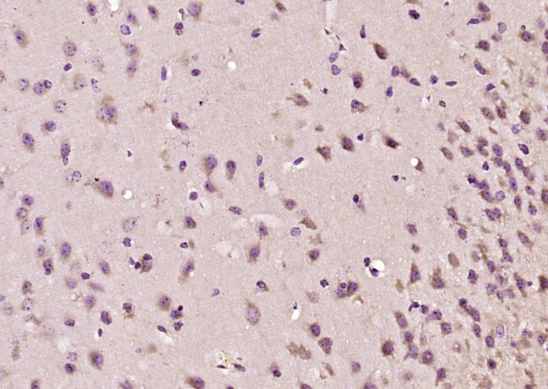
- 产品详情
- 实验流程
- 背景知识
Application 
| IHC-P, IHC-F, IF |
|---|---|
| Primary Accession | O60271 |
| Reactivity | Mouse |
| Predicted | Rat, Dog, Human, Chicken, Horse, Sheep |
| Host | Rabbit |
| Clonality | Polyclonal |
| Calculated MW | 146205 Da |
| Physical State | Liquid |
| Immunogen | KLH conjugated synthetic peptide derived from human JLP/JIP-4 |
| Epitope Specificity | 101-188/1321 |
| Isotype | IgG |
| Purity | affinity purified by Protein A |
| Buffer | 0.01M TBS (pH7.4) with 1% BSA, 0.02% Proclin300 and 50% Glycerol. |
| SUBCELLULAR LOCATION | Cytoplasm. Cytoplasm, perinuclear region. Note: Perinuclear distribution in response to stress signals such as UV radiation. |
| Important Note | This product as supplied is intended for research use only, not for use in human, therapeutic or diagnostic applications. |
| Background Descriptions | JIP-4 is a 1,321 amino acid protein encoded by the human gene SPAG9. It contains a large N-terminal extracellular domain, a short transmembrane helical domain, and a cytoplasmic domain. There are 6 N-glycosylation sites, several phosphorylation sites for cAMP/cGMP-dependent protein kinase, protein kinase C, and casein kinase II, and 10 putative myristoylation sites. There is also a leucine zipper motif, with 6 leucine repeats, that may aid in dimerization since there is no upstream basic domain characteristic of DNA binding proteins. The JNK-interacting protein (JIP) group of scaffold proteins selectively mediates JNK signaling by aggregating specific components of the MAPK cascade to form a functional JNK signaling module. JIP-4 is a cytoplasmic, perinuclear protein that has eight known isoforms whose expression varies by tissue and disease state. |
| Gene ID | 9043 |
|---|---|
| Other Names | C-Jun-amino-terminal kinase-interacting protein 4, JIP-4, JNK-interacting protein 4, Cancer/testis antigen 89, CT89, Human lung cancer oncogene 6 protein, HLC-6, JNK-associated leucine-zipper protein, JLP, Mitogen-activated protein kinase 8-interacting protein 4, Proliferation-inducing protein 6, Protein highly expressed in testis, PHET, Sperm surface protein, Sperm-associated antigen 9, Sperm-specific protein, Sunday driver 1, SPAG9 (HGNC:14524) |
| Dilution | IHC-P=1:100-500,IHC-F=1:100-500,IF=1:100-500 |
| Storage | Store at -20 °C for one year. Avoid repeated freeze/thaw cycles. When reconstituted in sterile pH 7.4 0.01M PBS or diluent of antibody the antibody is stable for at least two weeks at 2-4 °C. |
For Research Use Only. Not For Use In Diagnostic Procedures.
| Name | SPAG9 (HGNC:14524) |
|---|---|
| Function | The JNK-interacting protein (JIP) group of scaffold proteins selectively mediates JNK signaling by aggregating specific components of the MAPK cascade to form a functional JNK signaling module (PubMed:14743216). Regulates lysosomal positioning by acting as an adapter protein which links PIP4P1-positive lysosomes to the dynein- dynactin complex (PubMed:29146937). Assists PIKFYVE selective functionality in microtubule-based endosome-to-TGN trafficking (By similarity). |
| Cellular Location | Cytoplasm {ECO:0000250|UniProtKB:Q58A65}. Cytoplasm, perinuclear region {ECO:0000250|UniProtKB:Q58A65}. Lysosome membrane. Note=Perinuclear distribution in response to stress signals such as UV radiation {ECO:0000250|UniProtKB:Q58A65} |
| Tissue Location | Expressed only in testis on the round spermatids of stage I, II and II. Absent in spermatogonia and spermatocyte [Isoform 3]: Expressed in testis. |
Research Areas
Application Protocols
Provided below are standard protocols that you may find useful for product applications.
BACKGROUND
This product as supplied is intended for research use only, not for use in human, therapeutic or diagnostic applications.

终于等到您。ABCEPTA(百远生物)抗体产品。
点击下方“我要评价 ”按钮提交您的反馈信息,您的反馈和评价是我们最宝贵的财富之一,
我们将在1-3个工作日内处理您的反馈信息。
如有疑问,联系:0512-88856768 tech-china@abcepta.com.
¥ 1,250.00
Cat# AP54804


