


 癌症的基本特征包括细胞增殖、血管生成、迁移、凋亡逃避机制和细胞永生等。找到癌症发生过程中这些通路的关键标记物和对应的抗体用于检测至关重要。
 为您推荐一个泛素化位点预测神器——泛素化分析工具,可以为您的蛋白的泛素化位点作出预测和评分。
 细胞自噬受体图形绘图工具为你的蛋白的细胞受体结合位点作出预测和评分,识别结合到自噬通路中的蛋白是非常重要的,便于让我们理解自噬在正常生理、病理过程中的作用,如发育、细胞分化、神经退化性疾病、压力条件下、感染和癌症。
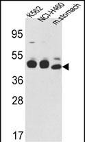
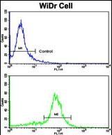
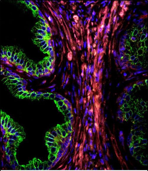
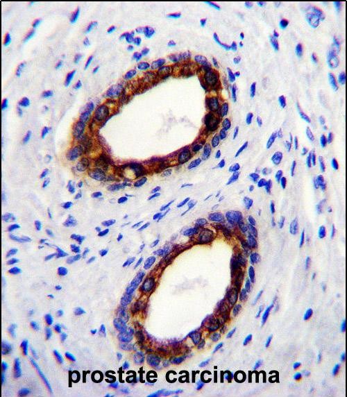
CYK18 Antibody (C-term)
Purified Rabbit Polyclonal Antibody (Pab)
- 产品详情
- 实验流程
- 背景知识
Application
| WB, FC, IF, IHC-P, E |
|---|---|
| Primary Accession | P05783 |
| Reactivity | Human, Rat, Mouse |
| Host | Rabbit |
| Clonality | Polyclonal |
| Isotype | Rabbit IgG |
| Calculated MW | 48058 Da |
| Antigen Region | 401-430 aa |
| Gene ID | 3875 |
|---|---|
| Other Names | Keratin, type I cytoskeletal 18, Cell proliferation-inducing gene 46 protein, Cytokeratin-18, CK-18, Keratin-18, K18, KRT18, CYK18 |
| Target/Specificity | This CYK18 antibody is generated from rabbits immunized with a KLH conjugated synthetic peptide between 401-430 amino acids from the C-terminal region of human CYK18. |
| Dilution | WB~~1:1000 FC~~1:10~50 IF~~1:10~50 IHC-P~~1:100~500 E~~Use at an assay dependent concentration. |
| Format | Purified polyclonal antibody supplied in PBS with 0.09% (W/V) sodium azide. This antibody is prepared by Saturated Ammonium Sulfate (SAS) precipitation followed by dialysis against PBS. |
| Storage | Maintain refrigerated at 2-8°C for up to 2 weeks. For long term storage store at -20°C in small aliquots to prevent freeze-thaw cycles. |
| Precautions | CYK18 Antibody (C-term) is for research use only and not for use in diagnostic or therapeutic procedures. |
| Name | KRT18 (HGNC:6430) |
|---|---|
| Synonyms | CYK18 |
| Function | Required for the formation of KRT8/KRT18 filaments that are involved in ARHGEF40-mediated actin stress fiber formation and tensional force-induced stress fiber formation and reinforcement (PubMed:26823019). Also acts downstream of ROCK kinase activation as part of a positive feedback mechanism in response to cellular mechanical stress loading (PubMed:26823019). Organization and orientation of KRT18 filaments are responsible for the properly elongated morphology of epithelial tubules (By similarity). Involved in the uptake of thrombin-antithrombin complexes by hepatic cells (By similarity). When phosphorylated, plays a role in filament reorganization. Involved in the delivery of mutated CFTR to the plasma membrane. Together with KRT8, is involved in interleukin-6 (IL-6)- mediated barrier protection. |
| Cellular Location | Nucleus matrix {ECO:0000250|UniProtKB:Q5BJY9}. Cytoplasm, perinuclear region. Nucleus, nucleolus. Cytoplasm {ECO:0000250|UniProtKB:Q5BJY9} |
| Tissue Location | Expressed in colon, placenta, liver and very weakly in exocervix. Increased expression observed in lymph nodes of breast carcinoma. |
For Research Use Only. Not For Use In Diagnostic Procedures.
Provided below are standard protocols that you may find useful for product applications.
BACKGROUND
KRT18 is the type I intermediate filament chain keratin 18. Keratin 18, together with its filament partner keratin 8, are perhaps the most commonly found members of the intermediate filament family. They are expressed in single layer epithelial tissues of the body. Mutations in its gene have been linked to cryptogenic cirrhosis.
REFERENCES
Zhang,Q., Clin. Cancer Res. 15 (10), 3557-3567 (2009)
Kruse,R., Folia Histochem. Cytobiol. 47 (1), 127-130 (2009)
Toivola,D.M., Hepatology 40 (2), 459-466 (2004)
终于等到您。ABCEPTA(百远生物)抗体产品。
点击下方“我要评价 ”按钮提交您的反馈信息,您的反馈和评价是我们最宝贵的财富之一,
我们将在1-3个工作日内处理您的反馈信息。
如有疑问,联系:0512-88856768 tech-china@abcepta.com.


