


 癌症的基本特征包括细胞增殖、血管生成、迁移、凋亡逃避机制和细胞永生等。找到癌症发生过程中这些通路的关键标记物和对应的抗体用于检测至关重要。
 为您推荐一个泛素化位点预测神器——泛素化分析工具,可以为您的蛋白的泛素化位点作出预测和评分。
 细胞自噬受体图形绘图工具为你的蛋白的细胞受体结合位点作出预测和评分,识别结合到自噬通路中的蛋白是非常重要的,便于让我们理解自噬在正常生理、病理过程中的作用,如发育、细胞分化、神经退化性疾病、压力条件下、感染和癌症。
PINK1 Antibody
- 产品详情
- 实验流程
- 背景知识
Application
| WB, E |
|---|---|
| Primary Accession | Q9BXM7 |
| Other Accession | NP_115785, 14165272 |
| Reactivity | Human, Mouse, Rat |
| Host | Rabbit |
| Clonality | Polyclonal |
| Isotype | IgG |
| Calculated MW | 62769 Da |
| Concentration (mg/ml) | 1 mg/mL |
| Conjugate | Unconjugated |
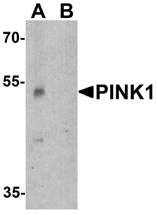
| Application Notes | PINK1 antibody can be used for detection of PINK1 by Western blot at 1 - 2 µg/ml. |
| Gene ID | 65018 |
|---|---|
| Other Names | Serine/threonine-protein kinase PINK1, mitochondrial, 2.7.11.1, BRPK, PTEN-induced putative kinase protein 1, PINK1 |
| Target/Specificity | PINK1; PINK1 antibody is human, mouse and rat reactive. At least two isoforms are known to exist; this antibody will only detect the longer isoform. PINK1 antibody will detect the cleaved and uncleaved form of PINK1. |
| Reconstitution & Storage | PINK1 antibody can be stored at 4℃ for three months and -20℃, stable for up to one year. |
| Precautions | PINK1 Antibody is for research use only and not for use in diagnostic or therapeutic procedures. |
| Name | PINK1 |
|---|---|
| Function | Serine/threonine-protein kinase which acts as a sensor of mitochondrial damage and protects against mitochondrial dysfunction during cellular stress. It phosphorylates mitochondrial proteins to coordinate mitochondrial quality control mechanisms that remove and replace dysfunctional mitochondrial components (PubMed:14607334, PubMed:15087508, PubMed:18443288, PubMed:18957282, PubMed:19229105, PubMed:19966284, PubMed:20404107, PubMed:20547144, PubMed:20798600, PubMed:22396657, PubMed:23620051, PubMed:23754282, PubMed:23933751, PubMed:24660806, PubMed:24751536, PubMed:24784582, PubMed:24896179, PubMed:24898855, PubMed:25527291, PubMed:32484300). Depending on the severity of mitochondrial damage, activity ranges from preventing apoptosis and stimulating mitochondrial biogenesis to eliminating severely damaged mitochondria via PINK1-PRKN-dependent mitophagy (PubMed:14607334, PubMed:15087508, PubMed:18443288, PubMed:19966284, PubMed:20404107, PubMed:20798600, PubMed:22396657, PubMed:23620051, PubMed:23933751, PubMed:24898855, PubMed:32047033, PubMed:32484300). When cellular stress results in irreversible mitochondrial damage, PINK1 accumulates at the outer mitochondrial membrane (OMM) where it phosphorylates pre-existing polyubiquitin chains at 'Ser-65', recruits PRKN from the cytosol to the OMM and activates PRKN by phosphorylation at 'Ser-65'; activated PRKN then ubiquinates VDAC1 and other OMM proteins to initiate mitophagy (PubMed:14607334, PubMed:15087508, PubMed:19966284, PubMed:20404107, PubMed:20798600, PubMed:23754282, PubMed:23933751, PubMed:24660806, PubMed:24751536, PubMed:24784582, PubMed:25474007, PubMed:25527291, PubMed:32047033). The PINK1-PRKN pathway also promotes fission of damaged mitochondria through phosphorylation and PRKN-dependent degradation of mitochondrial proteins involved in fission such as MFN2 (PubMed:18443288, PubMed:23620051, PubMed:24898855). This prevents the refusion of unhealthy mitochondria with the mitochondrial network or initiates mitochondrial fragmentation facilitating their later engulfment by autophagosomes (PubMed:18443288, PubMed:23620051). Also promotes mitochondrial fission independently of PRKN and ATG7-mediated mitophagy, via the phosphorylation and activation of DNM1L (PubMed:18443288, PubMed:32484300). Regulates motility of damaged mitochondria by promoting the ubiquitination and subsequent degradation of MIRO1 and MIRO2; in motor neurons, this likely inhibits mitochondrial intracellular anterograde transport along the axons which probably increases the chance of the mitochondria undergoing mitophagy in the soma (PubMed:22396657). Required for ubiquinone reduction by mitochondrial complex I by mediating phosphorylation of complex I subunit NDUFA10 (By similarity). Phosphorylates LETM1, positively regulating its mitochondrial calcium transport activity (PubMed:29123128). |
| Cellular Location | Mitochondrion outer membrane; Single-pass membrane protein. Mitochondrion inner membrane {ECO:0000250|UniProtKB:Q99MQ3}; Single-pass membrane protein. Cytoplasm, cytosol. Note=Localizes mostly in mitochondrion and the two smaller proteolytic processed fragments localize mainly in cytosol (PubMed:19229105). Upon mitochondrial membrane depolarization following damage, PINK1 import into the mitochondria is arrested, which induces its accumulation in the outer mitochondrial membrane, where it acquires kinase activity (PubMed:18957282) |
| Tissue Location | Highly expressed in heart, skeletal muscle and testis, and at lower levels in brain, placenta, liver, kidney, pancreas, prostate, ovary and small intestine. Present in the embryonic testis from an early stage of development |
For Research Use Only. Not For Use In Diagnostic Procedures.
Provided below are standard protocols that you may find useful for product applications.
BACKGROUND
The PTEN-induced putative kinase 1 (PINK1) is a serine/threonine protein kinase that localizes to mitochondria and is thought to protect cells from stress-induced mitochondrial dysfunction (reviewed in 1). PINK1 recruits the E3 ubiquitin ligase Parkin to mitochondria to initiate mitophagy, an autophagic process that clears damaged mitochondria within a cell (2). PINK1 is cleaved by the mitochondrial protease PARL (3). Mutations in this gene cause one form of autosomal recessive early-onset Parkinson disease (4).
REFERENCES
Matsuda S, Kitagishi Y, and Kobayashi M. Function and characteristics of PINK1 in mitochondria. Oxid. Med. Cell. Longev. 2013;601587.
Corti O, Lesage S, and Brice A. What genetics tells us about the causes and mechanism of Parkinson’s disease. Physiol. Rev. 2011; 91:1161-218.
Deas E, Plun-Favreau H, and Gandhi S, et al. PINK1 cleavage at position A103 by the mitochondrial protease PARL. Hum. Mol. Genet. 2011; 20:867-79.
Valente EM, Abou-Sleiman PM, Caputo V, et al. Hereditary early-onset Parkinson’s disease caused by mutations in PINK1. Science 2004; 304:1158-60.
终于等到您。ABCEPTA(百远生物)抗体产品。
点击下方“我要评价 ”按钮提交您的反馈信息,您的反馈和评价是我们最宝贵的财富之一,
我们将在1-3个工作日内处理您的反馈信息。
如有疑问,联系:0512-88856768 tech-china@abcepta.com.


